Tag Archive for: Chromosomes

Part 4: The story of chromosomes and inheritance

A Journey from Cork to Chromosomes

The Secret of Mitosis: How Scientists Discovered Cell Division


Chromosomes and the Secret of Inheritance

Walther Flemming had shown the precise and orderly sequence of cell division. But what were those mysterious threads? Why did they divide so carefully? And what did they carry?

The answers would come from a new generation of scientists at the turn of the 20th century.


Chromosomes Get Their Name

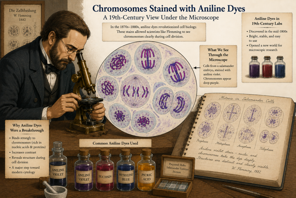
In 1888, a German anatomist named Heinrich Wilhelm Waldeyer gave the threads a new name: chromosomes.

  • “Chromo” means colour.
  • “Soma” means body.

Why? Because they picked up colour so vividly with the new dyes. The name stuck, and chromosomes became the stars of cell biology.

A Journey from Cork to Chromosomes
Figure 1: Chromosomes stained vividly with aniline dyes under a 19th-century microscope. (cartoon)

From Mitosis to Inheritance

Scientists now knew that chromosomes split evenly during mitosis. That meant daughter cells always received the same number. But another big mystery remained: How were traits passed from parents to offspring?

Gregor Mendel had already published his famous pea plant experiments back in 1866, showing that traits followed mathematical rules of inheritance.
Yet his work was forgotten for decades.

When it was rediscovered around 1900, scientists suddenly had two puzzles to connect:

  1. Mendel’s invisible units of heredity.
  2. The visible chromosomes that divided with such precision.

Could they be the same thing?


Theodor Boveri: A Careful Experimenter

The German biologist Theodor Boveri (1862–1915) was one of the first to see the connection.

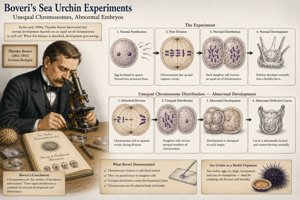
Working with sea urchin eggs, he performed delicate experiments. Sea urchins were perfect because their eggs were transparent, large, and divided rapidly. Boveri discovered something striking:

  • A cell could not develop properly unless it received a complete set of chromosomes.
  • If the distribution was abnormal if too many or too few chromosomes went into a cell, development failed.

This meant chromosomes were not just visible threads. They carried essential information needed for life.

A Journey from Cork to Chromosomes
Figure 2: Boveri’s sea urchin experiments — showing abnormal embryos when chromosomes were unevenly distributed. (cartoon)

Chromosomes as the Carriers of Heredity

Boveri concluded: chromosomes must be the physical carriers of heredity. Every single one mattered.
He was cautious but confident but the chromosomes were not identical; each had unique information. This was revolutionary. He had taken Flemming’s careful descriptions and added a powerful idea: chromosomes were not just for cell division, but also for inheritance of traits.


Walter Sutton and the Chromosome Theory

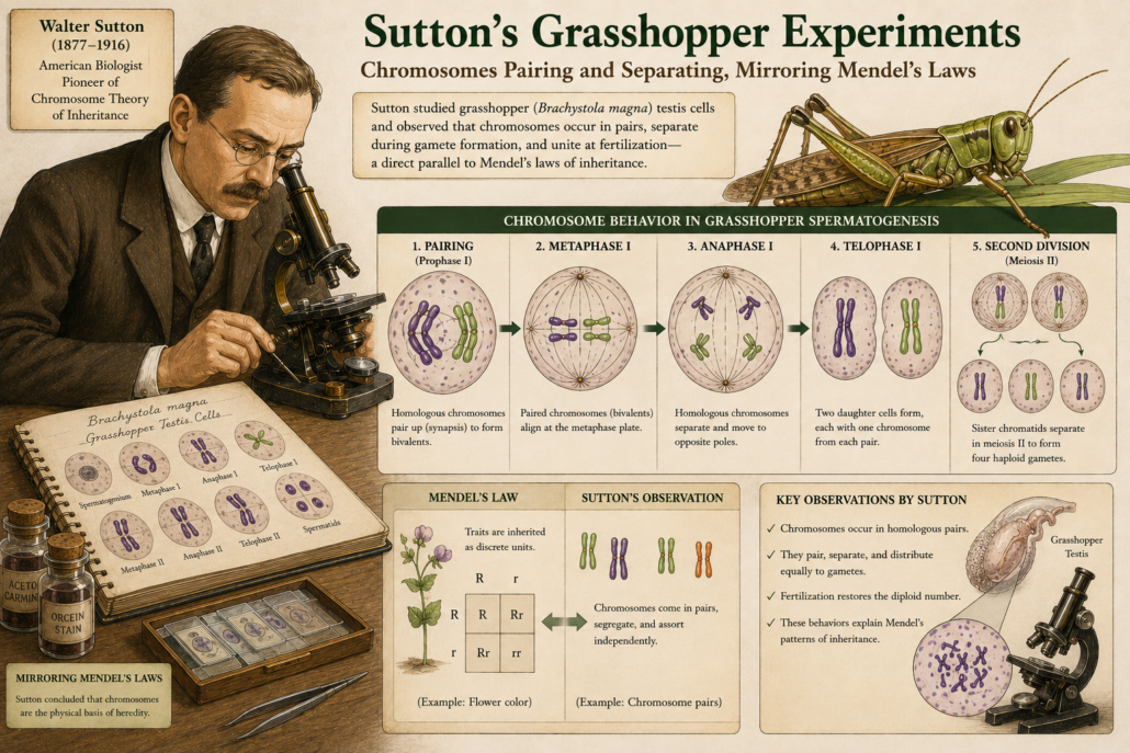
At the same time, across the Atlantic, a young American scientist named Walter Sutton was studying grasshopper cells. In 1902, Sutton noticed that chromosomes came in pairs. During cell division, each pair separated, with one chromosome going to each new cell. He realized this matched Mendel’s laws of inheritance:

  • Each parent contributes one factor (gene).
  • The factors separate and combine again in offspring.

Chromosomes behaved exactly like Mendel’s “hereditary units.”

A Journey from cork to chromosomes
Figure 3: Sutton’s grasshopper chromosomes pairing and separating, mirroring Mendel’s laws. (cartoon)

The Chromosome Theory of Inheritance

Together, Boveri and Sutton laid the foundation for the chromosome theory of inheritance.

It said:

  • Genes are located on chromosomes.
  • Chromosomes are passed from cell to cell, and from parent to offspring, through mitosis and meiosis.

This was the missing link between cell biology and genetics.


Why This Was a Turning Point

Up until Flemming, mitosis was just a strange cellular dance. With Boveri and Sutton, the meaning became clear:

  • Chromosomes were the carriers of heredity.
  • Mitosis ensured that every new cell received the full library of instructions.
  • Life’s continuity, from one cell to another, depended on this precise mechanism.

The mystery of cell division had transformed into the foundation of modern genetics.


A Legacy That Still Shapes Science

Today, we know chromosomes are made of DNA tightly wrapped around proteins.
We know they carry thousands of genes that code for every trait in living organisms.
But that knowledge rests on the careful experiments of Flemming, Boveri, Sutton, and their colleagues.

They took what seemed like meaningless threads and revealed them as the very carriers of life’s code.
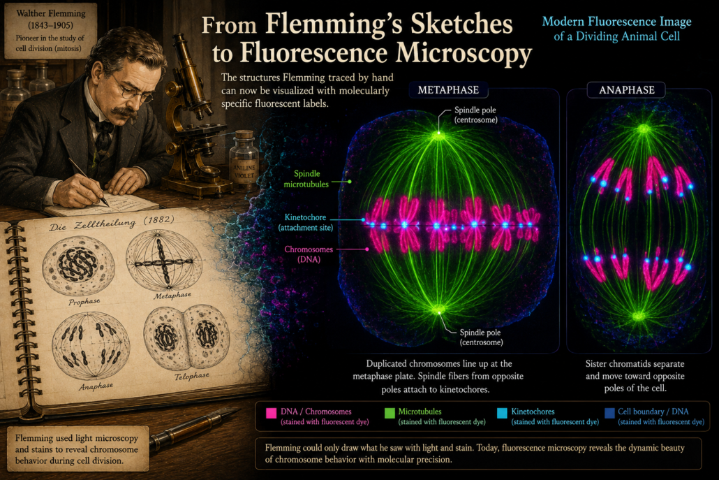

A Journey from Cork to Chromosomes
Figure 4: Modern fluorescence image of dividing chromosomes echoing Flemming’s hand-drawn sketches. (cartoon)

Part 5: Why Mitosis Matters Today, we will explore how this history connects to modern science: from cancer research to cloning, stem cells, and genetic medicine. Continue the journey…

Part 5: Why Mitosis Matters.
Let, connect the history to modern discoveries and why cell division still holds so many secrets.
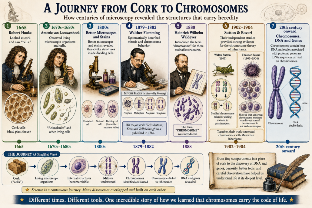

History of Cell Biology: From Hooke’s Cork Cells to Chromosomes


A Journey from Cork to Chromosomes

What if the story of genetics began with a piece of cork?
 Long before scientists understood DNA, genes, or chromosomes, they first had to discover cells and then learn what happened inside them.

Over several centuries, better microscopes, improved staining techniques, careful observations, and new ideas gradually transformed our understanding of life.

Robert Hooke looked at a thin slice of cork and saw tiny compartments, giving them the name “cells.” In the decades that followed, Antonie van Leeuwenhoek revealed a previously invisible world of microscopic organisms.

Robert Brown described the nucleus as a distinct structure in plant cells. Later, improved microscopy allowed scientists to observe the remarkable changes taking place as cells divided.

Matthias Schleiden and Theodor Schwann helped establish the idea that plants and animals are composed of cells.

Rudolf Virchow, building on earlier observations of cell

Robert Brown described the nucleus as a distinct structure in plant cells. Later, improved microscopy allowed scientists to observe the remarkable changes taking place as cells divided.

division, became closely associated with the principle omnis cellula e cellula that new cells arise from pre-existing cells.

Then the story became even more intriguing.

Scientists began seeing mysterious thread-like structures moving and changing during cell division. Walther Flemming systematically described their behaviour during mitosis, while Heinrich Wilhelm Waldeyer later introduced the term “chromosome.”

Finally, the independent work of Walter Sutton and Theodor Boveri provided important evidence linking chromosomes with heredity.

What began with a piece of cork was gradually becoming a much bigger story.


Timeline illustration of key discoveries in mitosis and chromosome theory.
A Journey from Cork to Chromosomes

A story about how life is organized.

A story about how cells reproduce.

And ultimately, a story about how biological information is passed from one generation to the next.

At the centre of this story was one extraordinary idea:

Cells come from cells.

The principle omnis cellula e cellula often translated as “every cell from a cell” became an important part of the developing cell theory. Virchow famously associated the principle with his cellular pathology work, although the understanding that cells arise through division was built on earlier observations, including the work of Robert Remak and others.

This is the journey from cork to chromosomes from simple observations under early microscopes to the foundations of modern cell biology and genetics.


What You’ll Discover in This Five-Part Journey


Part 1: The Birth of Cell Theory — From Robert Hooke’s Cork Cells to Virchow’s Principle

The journey begins in 1665, when Robert Hooke examined a thin slice of cork and saw a pattern of tiny box-like compartments.

He called them “cells.”

Hooke did not yet understand cells as the living units we know today. The cork tissue he observed was dead, and what he mainly saw were the empty spaces bounded by cell walls.

Over the following centuries, microscopes revealed an increasingly complex microscopic world. By 1838 and 1839, Matthias Schleiden and Theodor Schwann had helped establish the idea that plants and animals are composed of cells.

But one crucial question remained:

Where do new cells come from?

Observations of cell division including important work by scientists such as Robert Remak helped challenge the idea that cells could simply arise spontaneously. Virchow’s famous principle, omnis cellula e cellula, became closely associated with the idea that new cells arise from pre-existing cells.


The foundation of cell theory was taking shape.

→ Part 1 will explore:

Hooke → Leeuwenhoek → Brown → Schleiden → Schwann → Remak → Virchow


Part 2: The First Clues of Cell Division

During the 19th century, microscopes became more powerful, and new preparation and staining methods made internal cellular structures easier to observe.

Scientists began noticing something strange.

Inside dividing cells, thread-like material appeared to change shape and move in remarkably organized ways.

These were not random changes.

Something was happening inside the cell.

Researchers studying both plant and animal cells gradually accumulated evidence that cells reproduce through division and that structures within the nucleus undergo dramatic transformations during the process. Early work by scientists including Carl Nägeli, Eduard Strasburger, Friedrich Schneider, and others contributed to this growing picture, even though interpretations were not always correct.


A new mystery emerged:

What were these mysterious threads doing and why did they behave so precisely?

→ Part 2 leads directly to:

The mystery of dividing cells → the changing nuclear material → the search for the mechanism of cell division


Part 3: Walther Flemming and the Discovery of Mitosis

Walther Flemming took the investigation of cell division to a new level.

Using improved staining methods and meticulous microscopic observations, he followed the changing behaviour of nuclear material in dividing animal cells.

Flemming’s observations revealed an ordered sequence.

The material condensed.

It became organized.

It separated.

And it was distributed between the two resulting cells.

He systematically described the process that became known as mitosis, producing some of the earliest detailed accounts of chromosome behaviour during cell division. His major work, Zellsubstanz, Kern und Zelltheilung, was published in 1882.

Today, we describe the major stages as:

Prophase → Metaphase → Anaphase → Telophase


Flemming had revealed something fundamental:

Cell division was not chaos. It followed an astonishingly organized process.

Cell division was not chaos. It followed an astonishingly organized process.

But another mystery remained.

What was the purpose of these thread-like structures?

And could their precise behaviour somehow be connected to heredity?


Part 4: Chromosomes and the Mystery of Inheritance

By the late 19th century, the mysterious thread-like structures seen during cell division had become increasingly important.

In 1888, Heinrich Wilhelm Waldeyer introduced the term:

Chromosome.

Scientists could now give these remarkable structures a name.

But naming them did not explain what they did.

At the beginning of the 20th century, two seemingly separate areas of biology began to converge.

On one side was cell division and chromosome behaviour.

On the other was Mendelian inheritance.

Walter Sutton studied grasshopper chromosomes and noticed that their behaviour during meiosis closely matched the patterns predicted by Mendel’s laws of inheritance. Theodor Boveri’s work on chromosomes and development provided another important line of evidence. Together, their independent contributions helped support the emerging chromosome theory of inheritance.

Suddenly, the story became much bigger:

Cells + Chromosomes + Heredity


Chromosomes were no longer simply structures visible under a microscope.

They were increasingly suspected to be connected to the transmission of biological information from one generation to the next.

And that raised an even deeper question:

What were chromosomes actually made of?


Part 5: Why Mitosis Matters Today

The story does not end with Flemming’s microscope.

Today, the same process he painstakingly observed is studied using fluorescence microscopy, molecular biology, genetics, genome sequencing, and advanced imaging technologies.

Modern science has revealed that chromosomes contain long DNA molecules associated with proteins, and that genes are DNA sequences carried on chromosomes.

But mitosis is not simply a historical curiosity.

Cell division is fundamental to:

  • Growth
  • Development
  • Tissue repair
  • Stem cell biology
  • Embryonic developmentCancer research
  • Regenerative medicine

Every time a cell divides, an extraordinary challenge must be solved:

How can the cell accurately distribute its genetic material to the next generation of cells?

The same fundamental process that Flemming sketched by hand in the 19th century can now be observed with modern molecular tools.

From hand-drawn microscope sketches to fluorescent chromosomes.

From cork to chromosomes.

From chromosomes to DNA.

And from DNA to the genetic instructions that help shape every living organism.

The journey continues.

The microscope changed what humans could see.

Cell theory changed how we understood what we saw.

And chromosomes helped reveal how biological information could pass from one generation to the next.


What You’ll Discover in This Five-Part Journey

From Cork to Chromosomes — A Five-Part Journey

Part _Article

Part 1 _The Birth of Cell Theory

Part 2 _The First Clues of Cell Division

Part 3 _Walther Flemming and the Discovery of Mitosis

Part 4 _Chromosomes and Mystery of Inheritance

Part 5 _Why Mitosis Matters Today


What Can a Fruit Fly Teach Us About Ourselves? More Than You Think


The Tiny Genius That Changed Science: Drosophila melanogaster


The Little Fly With a Big Secret
You have probably seen it buzzing around your bananas — tiny, fast, and easy to miss. But did you know this little fly has helped scientists unlock some of the biggest mysteries in biology?

👉 Meet Drosophila melanogaster — the common fruit fly and the unexpected superstar of science!

This tiny insect may live for just a few weeks, but it’s made a massive impact. For over 100 years, scientists have studied fruit flies to discover how traits like eye colour, height, and even diseases are passed from parents to children. In fact, this bug was key to figuring out how genes work — long before we could even see DNA!

From your kitchen to the world’s top laboratories, the fruit fly has proven that even the smallest creatures can make the biggest difference.


A Riddle to Begin…

Let’s start with a riddle:

“Tiny wings, a simple fly,
Yet I helped science touch the sky.
With mutants, traits, and gene displays,
In my short life, I led the way.
What insect launched genetic fame?”

Answer: Drosophila melanogasterThe fly that taught humans about heredity!


Why Fruit Flies?

You might wonder: why study flies when we’re trying to understand human biology?

Well, it turns out that fruit flies are a perfect model organism for several reasons:

  • Short Life Cycle: A fruit fly grows from an egg to an adult in about 10 days. This means scientists can observe many generations quickly.
  • Simple Genome: Fruit flies have only 4 pairs of chromosomes, making it easier to study their DNA.
  • Easy to Keep in the Lab: They are small, don’t take up much space, and reproduce quickly — a dream for scientists!
  • Surprisingly Similar to Humans: Even though they’re tiny, fruit flies share about 60% of their genes with humans. Many of the genes that control development and disease in flies are similar to those in us.

The Beginning of Genetic Discovery

The real fame of Drosophila started in the early 1900s with a scientist named Thomas Hunt Morgan. He and his team at Columbia University began experimenting with fruit flies to understand how traits are passed from one generation to the next.

Here’s what they discovered:

  • Mutations Matter: By studying flies with white eyes (instead of the usual red), Morgan proved that genes are carried on chromosomes.
  • Sex-Linked Traits: He also showed that some traits are linked to sex chromosomes, which helped explain why some genetic diseases affect boys more than girls.
  • Gene Mapping: Morgan’s team created the first genetic maps, showing where genes are located on a chromosome.

For his ground breaking work, Thomas Hunt Morgan won the Nobel Prize in 1933. His research with Drosophila laid the foundation of modern genetics.


A Tiny Teacher in Modern Science

Even after more than 100 years, fruit flies are still teaching us new things!

Here’s what they’re helping us study today:

  • Brain and Behaviour: Scientists study how fruit fly brains control learning, memory, and sleep.
  • Disease Research: Fruit flies are used to model diseases like cancer, Parkinson’s, and Alzheimer’s — helping to test treatments.
  • Development: From a single cell to a full adult, fruit flies help us understand how living things grow and develop.

All of this is possible because we can control and observe mutations easily in fruit flies. Their transparent embryos, fast life cycle, and detailed genetic tools make them ideal for deep scientific exploration.


The Legacy of a Little Fly

From the kitchen counter to the Nobel stage, the journey of Drosophila melanogaster is nothing short of amazing. It has shown the world that even the smallest creatures can reveal life’s biggest secrets.

This tiny fly continues to help us answer questions like:

  • How do genes control growth?
  • Why do genetic diseases happen?
  • Can we fix or treat genetic disorders?

And many more…


Want to Explore More?

If you loved learning about this tiny genius, there’s so much more to explore in the world of genetics! From bacteria and worms to mice and zebrafish, science uses many amazing creatures to unlock the secrets of life.

Subscribe to our website for more fun and fascinating science facts.
Share this blog with fellow science lovers, and
Tell us in the comments — what other model organisms would you like to learn about next?


💡 Fun Fact:

A single female fruit fly can lay over 500 eggs in her lifetime. No wonder labs always have plenty of flies to study!