The Secret of Mitosis: How Scientists Discovered Cell Division
Why Mitosis Matters Today
The story of mitosis is not just history. It is a living science. Every day, billions of your cells divide through mitosis. Without it, growth, repair, and life itself would not be possible. But mitosis is more than just a biological fact. It is a key to medicine, biotechnology, and the future of human health.
Mitosis in Growth and Healing
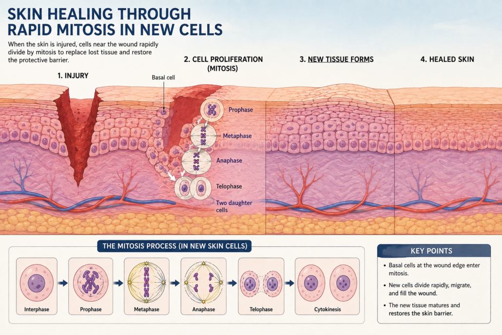
Think about a child growing taller. Or a cut on our skin closing as new tissue forms. Or the lining of our stomach renewing itself every few days.
All of these depend on mitosis. It ensures that each new cell is an exact copy of the one before. That continuity keeps tissues functioning, organs working, and bodies alive.
Figure 1: Animation still of skin healing through rapid mitosis in new cells.
When Mitosis Goes Wrong:
Cancer
But precision is crucial. If something goes wrong during mitosis, the results can be dangerous. Sometimes, a cell may divide uncontrollably. Instead of stopping when enough new cells are made, it keeps dividing. This is how cancer begins. Cancer is, in many ways, a disease of cell division. That is why scientists study mitosis so closely. Understanding the checkpoints, the signals, and the safeguards can lead to better treatments.
Chromosomes and Genetic Disorders
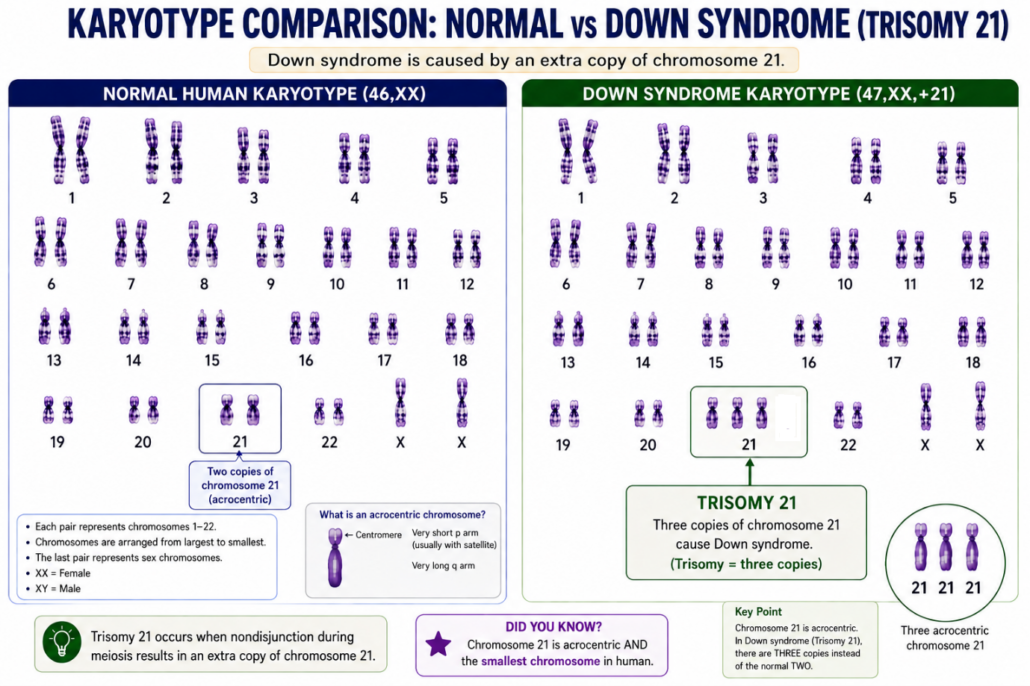
Mitosis also helps explain genetic disorders. If chromosomes are not copied or divided correctly, cells may end up with extra chromosomes or lack some. This can lead to developmental problems or diseases. For example:
Down syndrome happens when cells have an extra copy of chromosome 21.
Some disorders, like Cri-du-chat syndrome, happen when a small part of chromosome 5 is missing. Doctors have also found many other chromosomal disorders that occur when chromosomes are missing pieces or are rearranged.
Boveri’s insight that every chromosome matters are still guiding science today.
Figure 2: Karyotype image showing human chromosomes, including the extra one in Down syndrome.
Stem Cells and Regenerative Medicine
In modern labs, scientists harness mitosis in new ways. Stem cells are special because they can divide many times and become different types of cells.
In theory, they can repair damaged organs.
They can be used to grow tissues for transplants.
They could one day help cure diseases like Parkinson’s or heart failure.
All of this depends on the basic machinery of mitosis that is cells copying and dividing properly.
Mitosis and the Future of Genetics
The story also connects to DNA. In 1953, Watson and Crick revealed the double helix structure of DNA. That discovery explained what chromosomes are made of. Now we know genes lie along DNA, and chromosomes are their carriers. Every time a cell divides, mitosis ensures each new cell gets a complete copy of the genetic library. Today, with technologies like CRISPR gene editing, scientists can even change that library. But the properly copying and dividing lays the foundation of life’s continuity, still rests on mitosis.
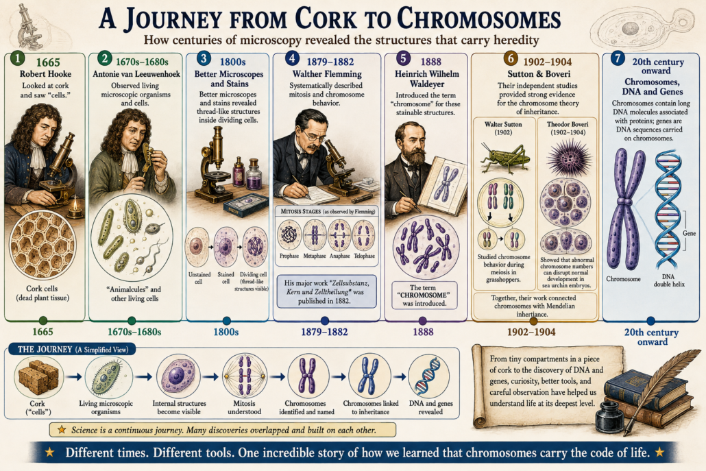
1665: Robert Hooke looked at cork and saw “cells.”
1670s: Antonie van Leeuwenhoek peered into living cells for the first time.
1800s: New dyes revealed thread-like structures inside dividing cells.
1879: Walther Flemming carefully described mitosis in his book Cell Substance, Nucleus, and Cell Division.
1888: Waldeyer named them “chromosomes.”
1902–1904: Boveri and Sutton showed chromosomes were essential for inheritance.
20th century onward: Chromosomes were revealed to be made of DNA, carrying genes.
It is a story of patience, of small steps, and of great leaps.
Figure 3: A Journey from Cork to Chromosomes– The Secret of Mitosis: How Scientists Discovered Cell Division
Why This Story Matters
The discovery of mitosis shows how science works. It is not the result of one genius moment. It is the outcome of centuries of curiosity, mistakes, improvements, and persistence.
From simple cork slices to high-powered microscopes, every discovery built on the last. Each scientist asked questions, saw something new, and passed the torch forward.
And today, when doctors fight cancer, or researchers grow tissues in a lab, or geneticists unlock the secrets of DNA, they are continuing that same journey.
Conclusion:
Mitosis is more than a cellular process. It is a story of life repeating itself, constantly, billions of times. It is the thread that connects us to our ancestors and carries us into the future. Every heartbeat, every breath, every moment somewhere inside us, mitosis is happening. The dance of the chromosomes goes on. And with it, the secret of life continues.
The process once observed through early microscopes is now central to understanding how organisms grow, tissues repair themselves, chromosomes are distributed, genetic disorders arise, and what can happen when normal cell-cycle control breaks down.
The Secret of Mitosis: How Scientists Discovered Cell Division
Chromosomes and the Secret of Inheritance
Walther Flemming had shown the precise and orderly sequence of cell division. But what were those mysterious threads? Why did they divide so carefully? And what did they carry?
The answers would come from a new generation of scientists at the turn of the 20th century.
Chromosomes Get Their Name
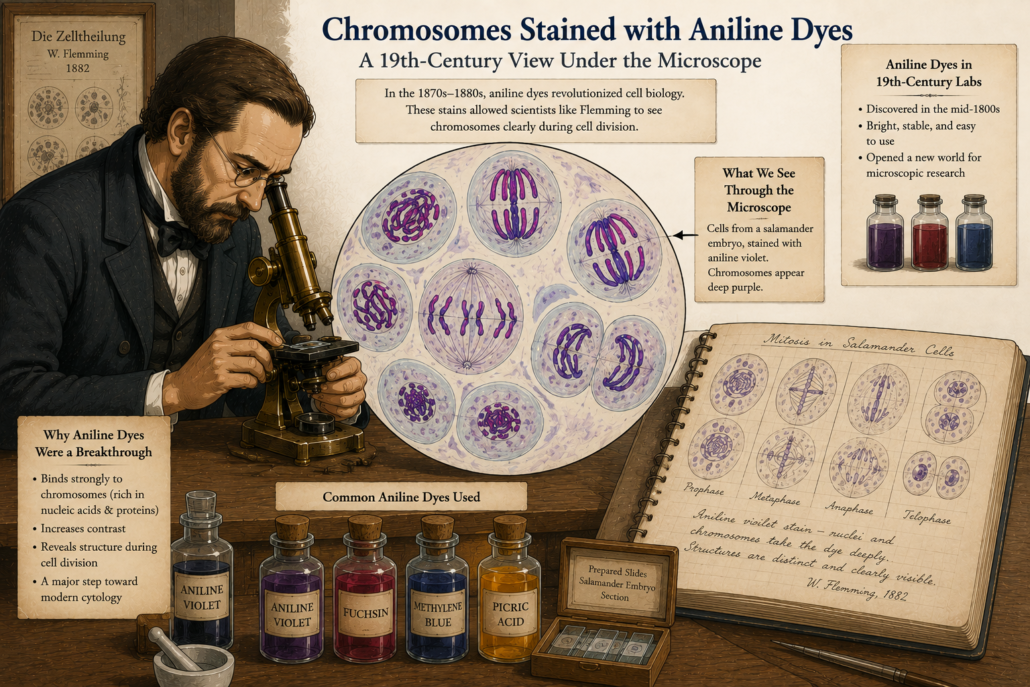
In 1888, a German anatomist named Heinrich Wilhelm Waldeyer gave the threads a new name: chromosomes.
“Chromo” means colour.
“Soma” means body.
Why? Because they picked up colour so vividly with the new dyes. The name stuck, and chromosomes became the stars of cell biology.
Figure 1: Chromosomes stained vividly with aniline dyes under a 19th-century microscope. (cartoon)
From Mitosis to Inheritance
Scientists now knew that chromosomes split evenly during mitosis. That meant daughter cells always received the same number. But another big mystery remained: How were traits passed from parents to offspring?
Gregor Mendel had already published his famous pea plant experiments back in 1866, showing that traits followed mathematical rules of inheritance. Yet his work was forgotten for decades.
When it was rediscovered around 1900, scientists suddenly had two puzzles to connect:
Mendel’s invisible units of heredity.
The visible chromosomes that divided with such precision.
Could they be the same thing?
Theodor Boveri: A Careful Experimenter
The German biologist Theodor Boveri (1862–1915) was one of the first to see the connection.
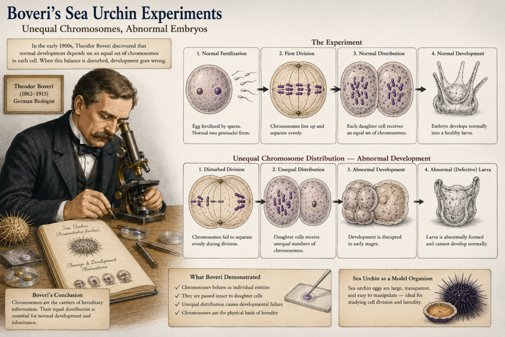
Working with sea urchin eggs, he performed delicate experiments. Sea urchins were perfect because their eggs were transparent, large, and divided rapidly. Boveri discovered something striking:
A cell could not develop properly unless it received a complete set of chromosomes.
If the distribution was abnormal if too many or too few chromosomes went into a cell, development failed.
This meant chromosomes were not just visible threads. They carried essential information needed for life.
Figure 2: Boveri’s sea urchin experiments — showing abnormal embryos when chromosomes were unevenly distributed. (cartoon)
Chromosomes as the Carriers of Heredity
Boveri concluded: chromosomes must be the physical carriers of heredity. Every single one mattered. He was cautious but confident but the chromosomes were not identical; each had unique information. This was revolutionary. He had taken Flemming’s careful descriptions and added a powerful idea: chromosomes were not just for cell division, but also for inheritance of traits.
Walter Sutton and the Chromosome Theory
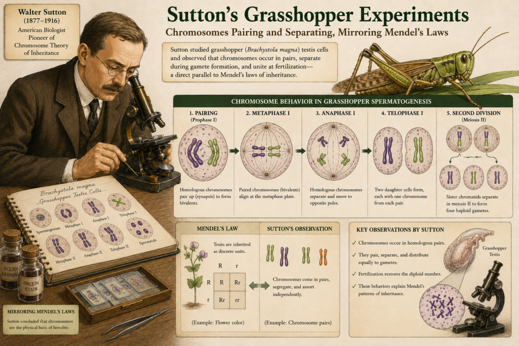
At the same time, across the Atlantic, a young American scientist named Walter Sutton was studying grasshopper cells. In 1902, Sutton noticed that chromosomes came in pairs. During cell division, each pair separated, with one chromosome going to each new cell. He realized this matched Mendel’s laws of inheritance:
Each parent contributes one factor (gene).
The factors separate and combine again in offspring.
Chromosomes behaved exactly like Mendel’s “hereditary units.”
Together, Boveri and Sutton laid the foundation for the chromosome theory of inheritance.
It said:
Genes are located on chromosomes.
Chromosomes are passed from cell to cell, and from parent to offspring, through mitosis and meiosis.
This was the missing link between cell biology and genetics.
Why This Was a Turning Point
Up until Flemming, mitosis was just a strange cellular dance. With Boveri and Sutton, the meaning became clear:
Chromosomes were the carriers of heredity.
Mitosis ensured that every new cell received the full library of instructions.
Life’s continuity, from one cell to another, depended on this precise mechanism.
The mystery of cell division had transformed into the foundation of modern genetics.
A Legacy That Still Shapes Science
Today, we know chromosomes are made of DNA tightly wrapped around proteins. We know they carry thousands of genes that code for every trait in living organisms. But that knowledge rests on the careful experiments of Flemming, Boveri, Sutton, and their colleagues.
They took what seemed like meaningless threads and revealed them as the very carriers of life’s code.
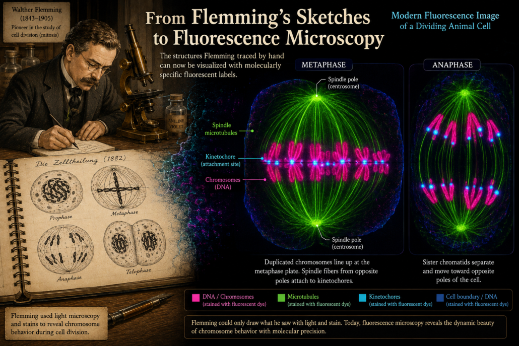
Figure 4: Modern fluorescence image of dividing chromosomes echoing Flemming’s hand-drawn sketches. (cartoon)
Part 5: Why Mitosis Matters Today, we will explore how this history connects to modern science: from cancer research to cloning, stem cells, and genetic medicine. Continue the journey…
Part 5: Why Mitosis Matters. Let, connect the history to modern discoveries and why cell division still holds so many secrets.
https://curiosciencequest.com/wp-content/uploads/2026/09/Part-4-Chromosomes-and-the-Mystery-of-Inheritance.png10241536info@curiosciencequest.comhttp://curiosciencequest.com/wp-content/uploads/2025/03/Untitled_design__1_-removebg-preview-1-300x300.pnginfo@curiosciencequest.com2026-09-04 12:31:422026-09-09 14:42:58Part 4: The story of chromosomes and inheritance
By the 1870s, scientists were catching glimpses of strange fibers inside the nucleus. Threads appeared, moved, and vanished during cell division. But no one had yet mapped the full sequence.
That changed with Walther Flemming.
The Scientist Behind the Discovery
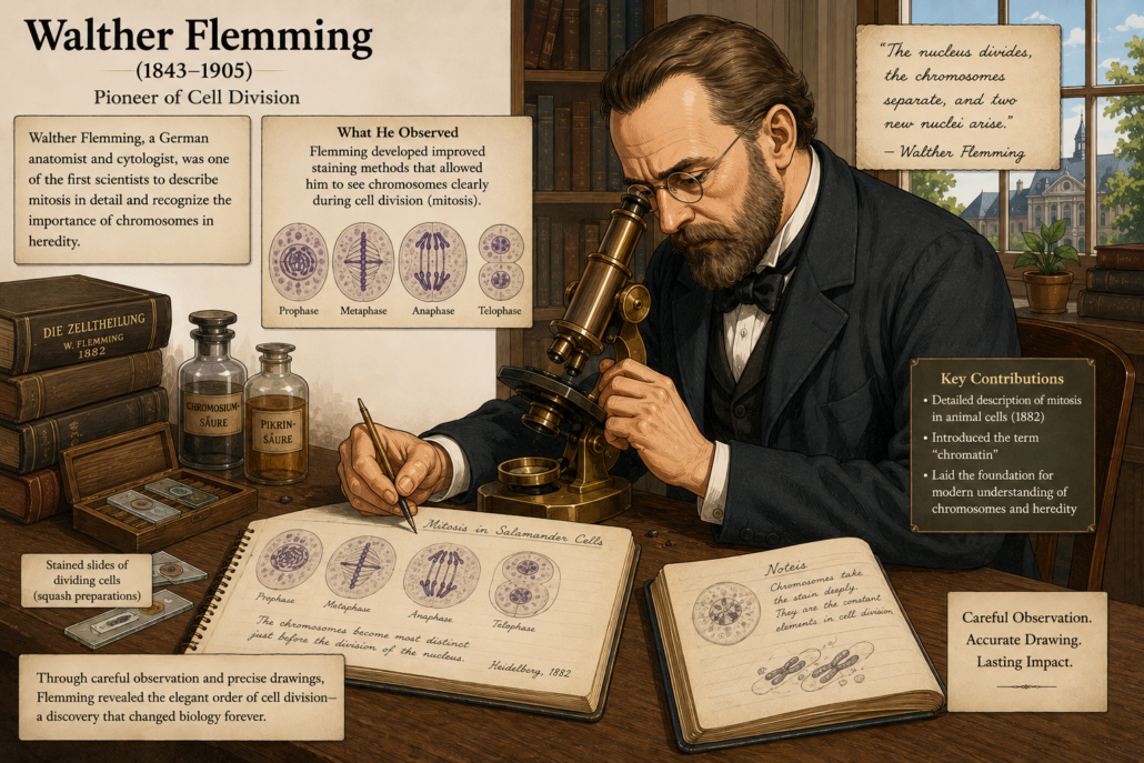
Walther Flemming (1843–1905) was a German anatomist and physician. He trained as a doctor but was drawn to microscopic anatomy the fine details of tissues and cells.
Unlike earlier observers, Flemming had access to better microscopes and a new set of tools: aniline dyes. These were synthetic colours developed from coal tar in the mid-19th century. They bound strongly to cell structures, especially the nucleus, and gave sharper, more consistent results than natural stains like carmine.
Flemming realized that with the right stains, he could follow the nucleus in fine detail as a cell divided.
Figure 1: Walther Flemming at his microscope, carefully sketching cell division. (cartoon)
A Patient Observer
Flemming spent years staring down the microscope at thin slices of tissue salamander embryos, corneal cells, and other transparent specimens where nuclei were easy to see.
He would watch patiently, draw what he saw, and compare hundreds of observations. Slowly, a clear pattern emerged. He saw that cell division was not random or disorderly, but it followed a precise, step-by-step process rather than occurring by chance.
The Stages of Mitosis
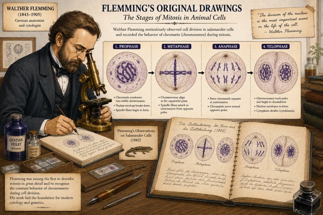
Flemming documented a series of stages that every dividing nucleus seemed to pass through:
Prophase – The nucleus, normally round and quiet, suddenly filled with long, dark threads. These threads shortened and thickened, becoming more distinct. (We now know these threads are chromosomes condensing.)
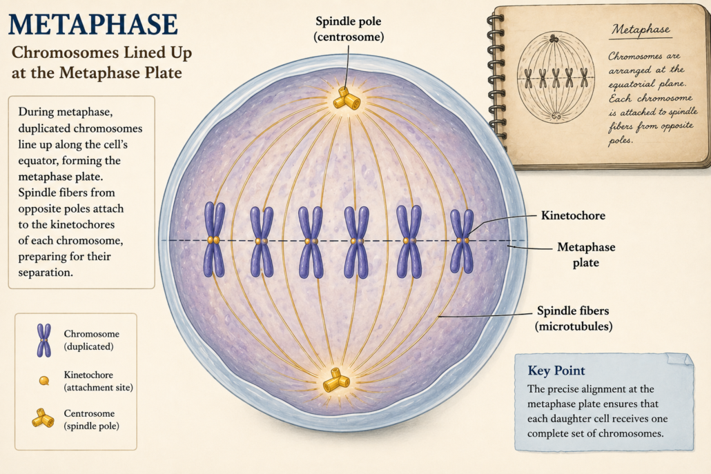
Metaphase – The threads arranged themselves in the middle of the cell, forming a striking plate-like structure. (We now call this the metaphase plate.)
Anaphase – The threads split lengthwise. Each half moved steadily to opposite ends of the cell. (This ensures each new cell receives the same set of chromosomes.)
Telophase – At the two ends, the threads disappeared again into a fine network, and new nuclei re-formed.
Finally, the whole cell itself divided, producing two daughter cells, each with a complete nucleus.
Figure 2: Flemming’s original drawings showing the stages of mitosis: prophase, metaphase, anaphase, and telophase. (cartoon)
Naming the Process
In 1882, Flemming published his great work, Cell Substance, Nucleus, and Cell Division.
In it, he coined the term “mitosis,” from the Greek word mitos, meaning “thread.” It was a perfect description: division looked like a dance of threads inside the cell.
Why It Was Revolutionary
Flemming had achieved something extraordinary. For the first time in history, scientists could see how one cell became two at the nuclear level.
He proved that division was not random but it followed a reproducible sequence.
He gave names and drawings that future scientists could use and refine.
He opened the door to understanding inheritance, because whatever these “threads” were, they clearly carried something important into daughter cells.
At the time, no one knew these threads were chromosomes containing DNA. That connection would come later. But Flemming’s careful descriptions laid the foundation.
A Turning Point: Metaphase
Metaphase was the most striking stage Flemming described. For the first time, scientists observed chromosomes aligning in a precise order. This was not random. It indicated an internal mechanism controlling their movement. Cell division was therefore not just a physical split but a highly organized biological process.
Figure 3: Chromosomes lined up at the metaphase plate.
Legacy of Flemming
Flemming did not live to see the discovery of DNA or the chromosome theory of inheritance. But without him, those later insights would not have been possible.
His drawings remain iconic even today. Modern fluorescent microscopy confirms almost exactly what he sketched by hand more than 140 years ago.
Walther Flemming showed the world the secret arrangement inside the cell. He gave us the language, the stages, and the concept of mitosis.
Part 4: Chromosomes and Inheritance, we will explore how scientists like Theodor Boveri and Walter Sutton realized that chromosomes were not just threads but they were the very carriers of heredity. Continue the journey…
Part 4 The story of chromosomes and inheritance. This is where mitosis connects to heredity and the secret of life itself.
https://curiosciencequest.com/wp-content/uploads/2026/09/Part-3Walther-Flemming-and-the-Discovery-of-Mitosis.png9411672info@curiosciencequest.comhttp://curiosciencequest.com/wp-content/uploads/2025/03/Untitled_design__1_-removebg-preview-1-300x300.pnginfo@curiosciencequest.com2026-09-03 09:31:122026-09-09 14:39:37Part 3: Walther Flemming and the Discovery of Mitosis
The Secret of Mitosis: How Scientists Discovered Cell Division
By the mid-1800s, the microscope had become the scientist’s window into life. But looking inside a living cell was still very difficult. Cells were tiny, transparent, and constantly moving.
To solve this, scientists began experimenting with dyes and stains. Coloured chemicals could bind to certain parts of a cell, making invisible structures visible. This simple trick changed biology.
Carmine and the First Threads
One of the most useful dyes was carmine, a deep red pigment extracted from insects. When applied to cells, it stained the dense material inside the nucleus more strongly than the rest of the cell.
In the 1840s and 1850s, botanists such as Carl Nageli and Hugo von Mohl began to describe “thread-like” forms in plant cell nuclei. They didn’t fully understand what they were looking at, but their drawings showed thin, elongated fibers that seemed to appear during cell division.
These threads were, in fact, chromosomes though that word did not yet exist.
Figure 1: Early drawings by Nageli showing thread-like structures in dividing plant cells. (cartoon)
Robert Remak and Rudolf Virchow
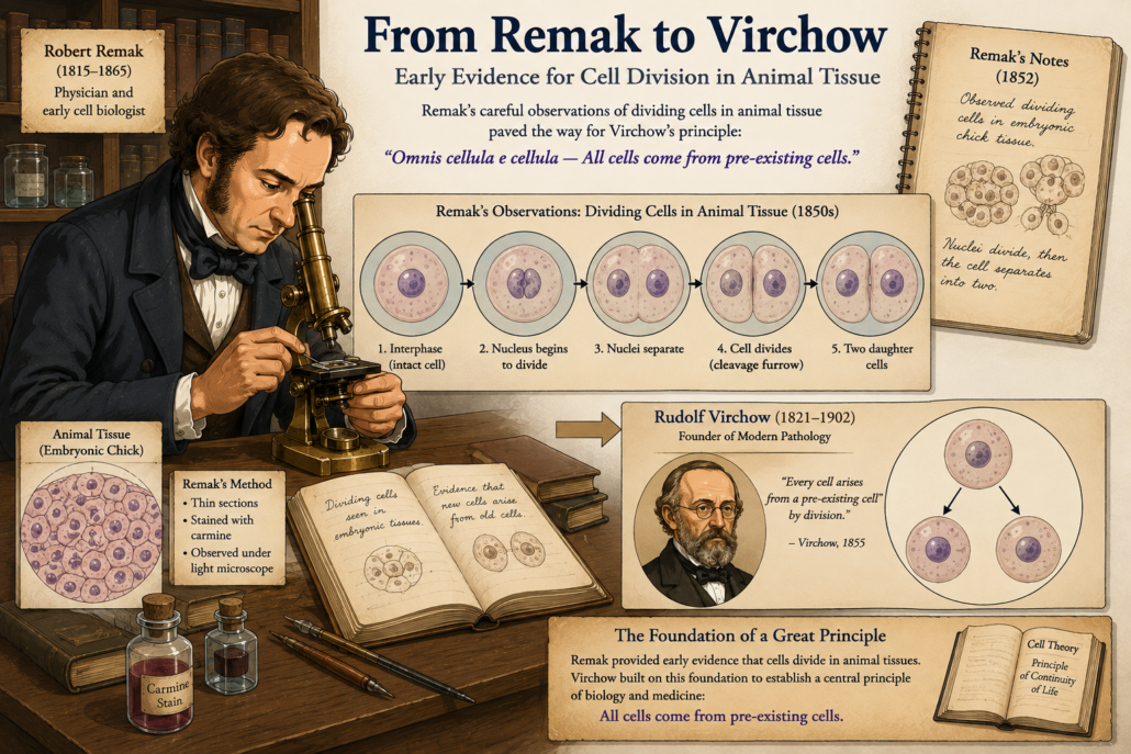
Meanwhile, in 1841, Robert Remak, a young German physician, was studying the development of chicken embryos. Using careful observation and early staining, he noticed that new cells did not appear spontaneously. Instead, they formed by binary division that is one cell splitting into two.
This was one of the earliest direct confirmations of Virchow’s later principle: cells arise from cells.
But Remak’s work was not widely accepted at first. It was Virchow, a more influential figure, who popularized the idea in 1855 with his famous phrase “Omnis cellula e cellula.”
Still, Remak deserves credit for being among the first to carefully document cell division in animal tissue.
Figure 2: Early evidence for cell division in animal tissue Remak’s careful work paved the way for Virchow’s principle that all cells come from pre-existing cells.
Strasburger and the Plants
By the 1870s, techniques had advanced further. German botanist Eduard Strasburger used stronger microscopes and refined stains to study plant cells, especially in lilies and algae.
He provided detailed descriptions of the nucleus dividing into two during cell reproduction. He also noticed that the thread-like forms which we now recognize as chromosomes moved in a regular, organized way.
Strasburger’s observations hinted at a precise, step-by-step process. Division was not random. It followed an orderly sequence.
Figure 3 By the 1870s, Strasburger’s refined plant studies and Bütschli’s animal research revealed the universal ‘threads’ of life early glimpses of what we now call chromosomes.
Butschli and the “Nuclear Threads”
Another important contributor was Otto Butschli, who in the 1870s studied the eggs of worms and other simple animals. His work provided evidence that the “threads” were universal not just in plants, but in animals too.
This was a crucial step. The mysterious fibers inside cells were not accidents or artifacts of staining. They were real biological structures, present in all dividing cells.
By now, scientists were seeing glimpses of chromosomes, though they did not yet understand their true importance.
The Stage Is Set
By the late 19th century, one thing was clear: cells divided by a process involving the nucleus. Inside the nucleus, thread-like structures appeared, lined up, separated, and disappeared again.
But who would finally put the pieces together into a complete, step-by-step description?
That task would fall to a German anatomist named Walther Flemming.
Part 3: Walther Flemming and the Birth of “Mitosis”, we will explore how Flemming, using aniline dyes and patient observation, became the first person to clearly describe the entire process of mitosis. Continue the journey…
https://curiosciencequest.com/wp-content/uploads/2026/09/Part-2-The-First-Clues-of-Cell-Division.png9411672info@curiosciencequest.comhttp://curiosciencequest.com/wp-content/uploads/2025/03/Untitled_design__1_-removebg-preview-1-300x300.pnginfo@curiosciencequest.com2026-09-02 16:47:022026-09-09 14:30:47Part 2:The First Clues of Cell Division
History of Cell Biology: From Hooke’s Cork Cells to Chromosomes
From Cork to Cells: Robert Hooke and the Birth of Cell Biology
What is the history of cell biology?
The history of cell biology began with Robert Hooke’s observation of cork cells in 1665 and progressed through the discoveries of Leeuwenhoek, Brown, Schleiden, schwann, Virchow and Flemming.
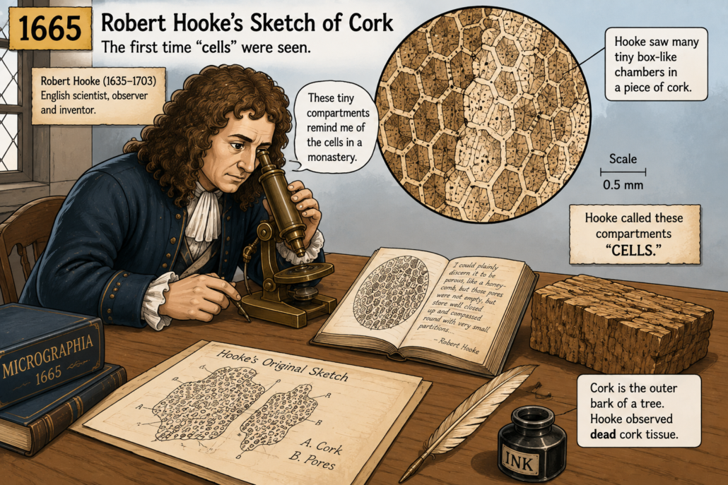
Imagine peeling the bark from a tree and placing a thin slice under a primitive microscope. That is exactly what happened in 1665, when the English scientist Robert Hooke looked at cork. Through his hand-built compound microscope, he saw something no one had described before: tiny, box-like compartments.
Hooke thought they looked like the small rooms where monks lived. He called them “cells.”
This was the first recorded use of the word “cell” in biology. Of course, Hooke was not actually seeing living cells. He was looking at the empty walls of dead plant tissue. The living contents had long dried away, leaving behind only the rigid outlines.
But his observation was revolutionary. Hooke’s book Micrographia (1665) inspired curiosity across Europe. The idea that nature could be studied with magnifying lenses became a serious pursuit.
Figure 1: Robert Hooke’s 1665 sketch of cork, showing tiny “cells.”showing tiny “cells.”
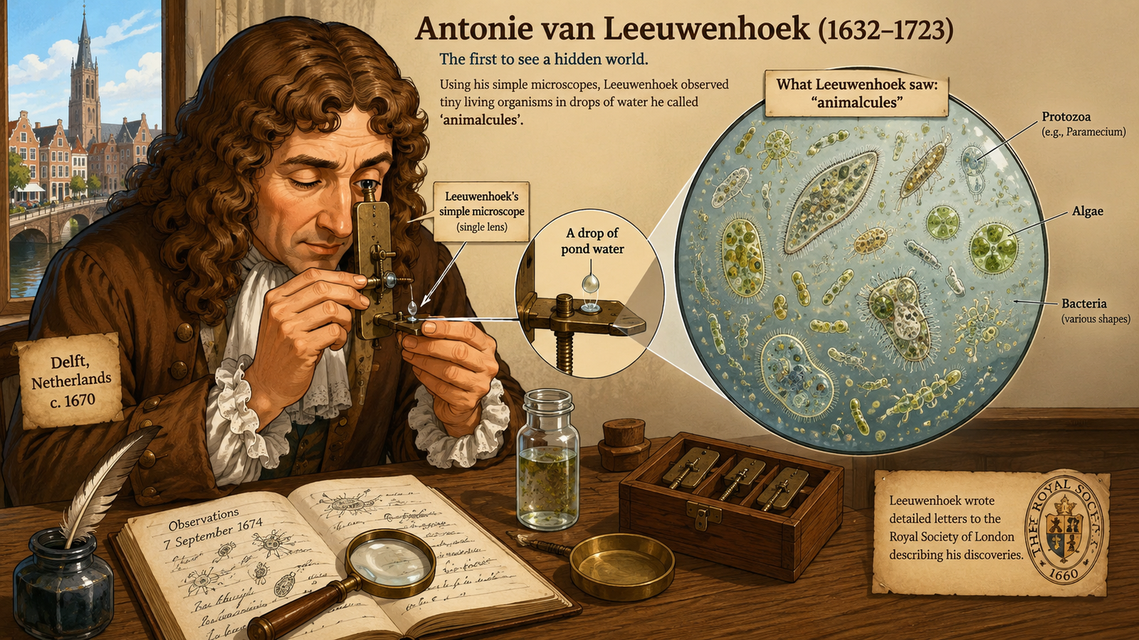
At the same time, another pioneer was grinding glass into lenses that were far better than Hooke’s. This man was Antonie van Leeuwenhoek, a Dutch tradesman with a passion for magnification. His microscopes were small, single-lens devices, but they were powerful
Leeuwenhoek used his lenses to look at everything he could imagine from pond water, blood, semen, even scrapings from his own teeth. What he discovered amazed the world. He described tiny moving creatures that no one had seen before and named them ‘animalcules.
These were the first recorded observations of living microorganisms, bacteria, protozoa, and even sperm cells.
Figure 2: Antonie van Leeuwenhoek’s simple microscope and his “animalcules” in water droplets.
For the first time, people began to understand that life was built from tiny, hidden structures. But there was still no single theory to explain it all. Hooke had described cells in plants, and Leeuwenhoek had observed animal and bacterial forms. The connection between them remained unclear.
More than 150 years would pass before scientists connected these dots.
The Nucleus Appears
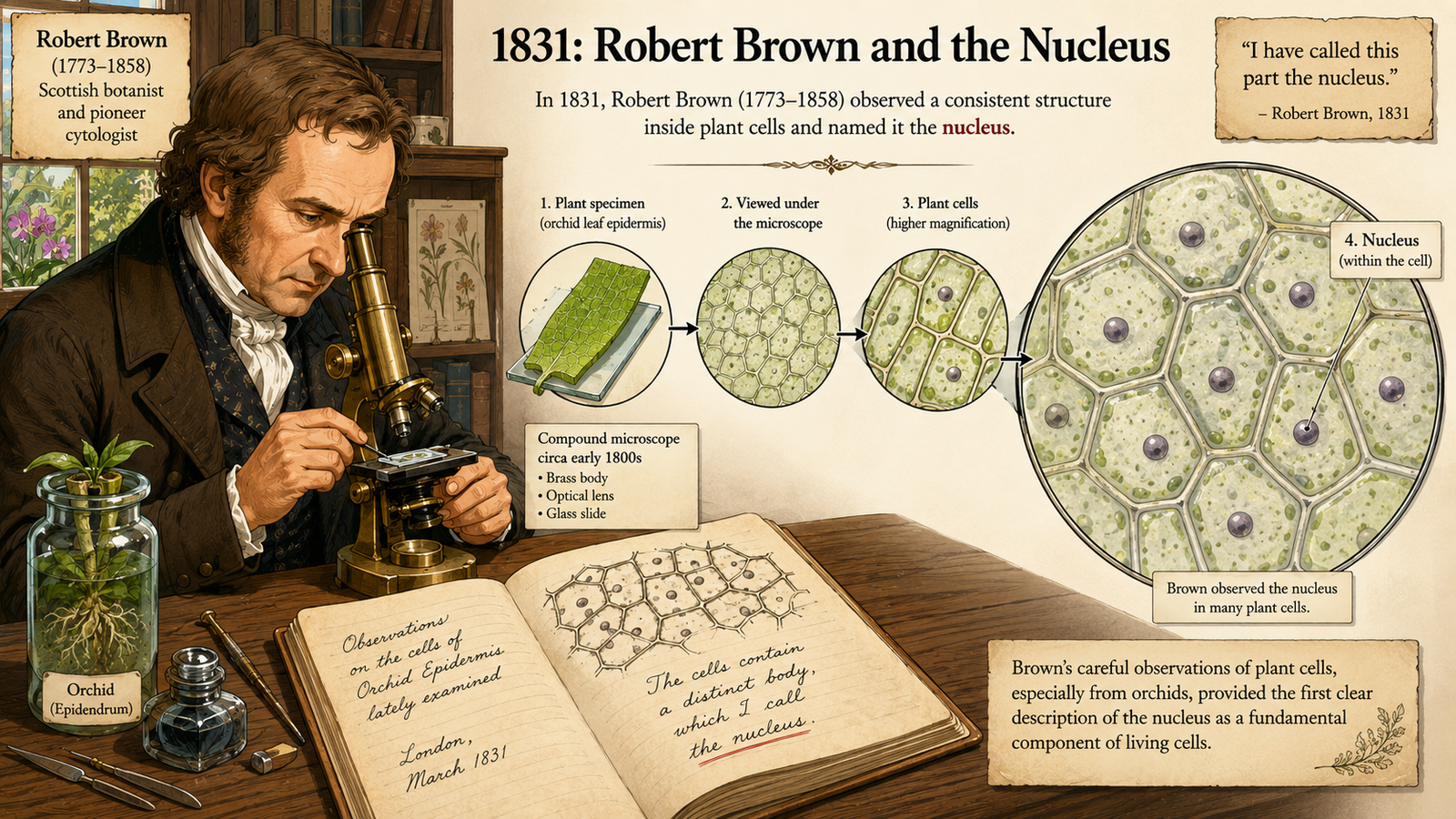
In 1831, Scottish botanist Robert Brown was studying orchids. While observing the cells of these plants, he noticed a consistent, dark, rounded body inside each one. He named it the nucleus.
This was a turning point. Cells were not just empty spaces surrounded by walls. They had internal parts. They had organization. The nucleus seemed to be a central structure, and later it would prove essential for heredity.
Figure 3: Robert Brown’s 1831 drawing of plant cells, highlighting the nucleus.
The Cell Theory
By the 1830s and 1840s, microscopes were improving. Glassmaking had advanced, and staining methods with dyes like carmine and iodine made cellular details easier to see.
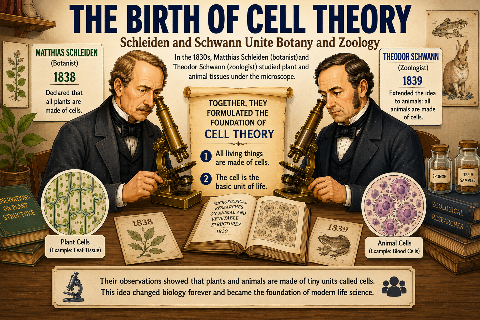
In Europe, two scientists named Matthias Schleiden (a botanist) and Theodor Schwann (a zoologist) began studying plant and animal tissues under the microscope.
In 1838, Schleiden declared that all plants are made of cells. In 1839, Schwann extended the idea to animals. Together, they laid down the foundation of what became known as the Cell Theory:
All living things are made of cells.
The cell is the basic unit of life
Figure 4: Schielden and Schwann unite Botany and Zoology
This was revolutionary. Suddenly, plants, animals, and even humans were united by a common building block.
Yet one mystery remained: Where do new cells come from?
At the time, many scientists still believed in “spontaneous generation” the idea that life could appear out of non-living matter. Some thought cells could simply crystallize or form a new inside organisms.
It was not until 1855 that German physician Rudolf Virchow delivered the famous phrase: “Omnis cellula e cellula.”Every cell comes from another cell.
This simple idea challenged the long-held belief that cells could arise by spontaneous generation. It showed cells, could not appear from nothing but they had to come from the division of pre-existing cells.
Virchow’s principle would become the third pillar of the Cell Theory.
A Hidden Process
But here lay the puzzle: How exactly does one cell become two?
Scientists had no clear picture of the process. They could sometimes glimpse strange, thread-like structures inside dividing cells, especially when they used new stains. But these observations were brief and confusing.
The stage was set for a discovery that would change biology forever the discovery of mitosis, the process of cell division.
Part 2: The First Clues of Cell Division, we will explore the 1800s when new dyes and microscopes revealed mysterious threads inside cells, and Virchow’s principle met its first experimental tests. Continue the journey…..
The Secret of Mitosis: How Scientists Discovered Cell Division
https://curiosciencequest.com/wp-content/uploads/2026/08/blog_the-birth-of-cell-theory-e1787678814815.png6751013info@curiosciencequest.comhttp://curiosciencequest.com/wp-content/uploads/2025/03/Untitled_design__1_-removebg-preview-1-300x300.pnginfo@curiosciencequest.com2026-09-02 03:33:242026-09-11 11:30:46Part 1: The Birth of Cell Theory
What if the story of genetics began with a piece of cork? Long before scientists understood DNA, genes, or chromosomes, they first had to discover cells and then learn what happened inside them.
Over several centuries, better microscopes, improved staining techniques, careful observations, and new ideas gradually transformed our understanding of life.
Robert Hooke looked at a thin slice of cork and saw tiny compartments, giving them the name “cells.” In the decades that followed, Antonie van Leeuwenhoek revealed a previously invisible world of microscopic organisms.
Robert Brown described the nucleus as a distinct structure in plant cells. Later, improved microscopy allowed scientists to observe the remarkable changes taking place as cells divided.
Matthias Schleiden and Theodor Schwann helped establish the idea that plants and animals are composed of cells.
Rudolf Virchow, building on earlier observations of cell
Robert Brown described the nucleus as a distinct structure in plant cells. Later, improved microscopy allowed scientists to observe the remarkable changes taking place as cells divided.
division, became closely associated with the principle omnis cellula e cellula that new cells arise from pre-existing cells.
Then the story became even more intriguing.
Scientists began seeing mysterious thread-like structures moving and changing during cell division. Walther Flemming systematically described their behaviour during mitosis, while Heinrich Wilhelm Waldeyer later introduced the term “chromosome.”
Finally, the independent work of Walter Sutton and Theodor Boveri provided important evidence linking chromosomes with heredity.
What began with a piece of cork was gradually becoming a much bigger story.
A Journey from Cork to Chromosomes
A story about how life is organized.
A story about how cells reproduce.
And ultimately, a story about how biological information is passed from one generation to the next.
At the centre of this story was one extraordinary idea:
Cells come from cells.
The principle omnis cellula e cellula often translated as “every cell from a cell” became an important part of the developing cell theory. Virchow famously associated the principle with his cellular pathology work, although the understanding that cells arise through division was built on earlier observations, including the work of Robert Remak and others.
This is the journey from cork to chromosomes from simple observations under early microscopes to the foundations of modern cell biology and genetics.
What You’ll Discover in This Five-Part Journey
Part 1: The Birth of Cell Theory — From Robert Hooke’s Cork Cells to Virchow’s Principle
The journey begins in 1665, when Robert Hooke examined a thin slice of cork and saw a pattern of tiny box-like compartments.
He called them “cells.”
Hooke did not yet understand cells as the living units we know today. The cork tissue he observed was dead, and what he mainly saw were the empty spaces bounded by cell walls.
Over the following centuries, microscopes revealed an increasingly complex microscopic world. By 1838 and 1839, Matthias Schleiden and Theodor Schwann had helped establish the idea that plants and animals are composed of cells.
But one crucial question remained:
Where do new cells come from?
Observations of cell division including important work by scientists such as Robert Remak helped challenge the idea that cells could simply arise spontaneously. Virchow’s famous principle, omnis cellula e cellula, became closely associated with the idea that new cells arise from pre-existing cells.
During the 19th century, microscopes became more powerful, and new preparation and staining methods made internal cellular structures easier to observe.
Scientists began noticing something strange.
Inside dividing cells, thread-like material appeared to change shape and move in remarkably organized ways.
These were not random changes.
Something was happening inside the cell.
Researchers studying both plant and animal cells gradually accumulated evidence that cells reproduce through division and that structures within the nucleus undergo dramatic transformations during the process. Early work by scientists including Carl Nägeli, Eduard Strasburger, Friedrich Schneider, and others contributed to this growing picture, even though interpretations were not always correct.
A new mystery emerged:
What were these mysterious threads doing and why did they behave so precisely?
→ Part 2 leads directly to:
The mystery of dividing cells → the changing nuclear material → the search for the mechanism of cell division
Part 3: Walther Flemming and the Discovery of Mitosis
Walther Flemming took the investigation of cell division to a new level.
Using improved staining methods and meticulous microscopic observations, he followed the changing behaviour of nuclear material in dividing animal cells.
Flemming’s observations revealed an ordered sequence.
The material condensed.
It became organized.
It separated.
And it was distributed between the two resulting cells.
He systematically described the process that became known as mitosis, producing some of the earliest detailed accounts of chromosome behaviour during cell division. His major work, Zellsubstanz, Kern und Zelltheilung, was published in 1882.
Today, we describe the major stages as:
Prophase → Metaphase → Anaphase → Telophase
Flemming had revealed something fundamental:
Cell division was not chaos. It followed an astonishingly organized process.
Cell division was not chaos. It followed an astonishingly organized process.
But another mystery remained.
What was the purpose of these thread-like structures?
And could their precise behaviour somehow be connected to heredity?
Part 4: Chromosomes and the Mystery of Inheritance
By the late 19th century, the mysterious thread-like structures seen during cell division had become increasingly important.
In 1888, Heinrich Wilhelm Waldeyer introduced the term:
Chromosome.
Scientists could now give these remarkable structures a name.
But naming them did not explain what they did.
At the beginning of the 20th century, two seemingly separate areas of biology began to converge.
On one side was cell division and chromosome behaviour.
Walter Sutton studied grasshopper chromosomes and noticed that their behaviour during meiosis closely matched the patterns predicted by Mendel’s laws of inheritance. Theodor Boveri’s work on chromosomes and development provided another important line of evidence. Together, their independent contributions helped support the emerging chromosome theory of inheritance.
Suddenly, the story became much bigger:
Cells + Chromosomes + Heredity
Chromosomes were no longer simply structures visible under a microscope.
They were increasingly suspected to be connected to the transmission of biological information from one generation to the next.
And that raised an even deeper question:
What were chromosomes actually made of?
Part 5: Why Mitosis Matters Today
The story does not end with Flemming’s microscope.
Today, the same process he painstakingly observed is studied using fluorescence microscopy, molecular biology, genetics, genome sequencing, and advanced imaging technologies.
Modern science has revealed that chromosomes contain long DNA molecules associated with proteins, and that genes are DNA sequences carried on chromosomes.
But mitosis is not simply a historical curiosity.
Cell division is fundamental to:
Growth
Development
Tissue repair
Stem cell biology
Embryonic developmentCancer research
Regenerative medicine
Every time a cell divides, an extraordinary challenge must be solved:
How can the cell accurately distribute its genetic material to the next generation of cells?
The same fundamental process that Flemming sketched by hand in the 19th century can now be observed with modern molecular tools.
From hand-drawn microscope sketches to fluorescent chromosomes.
From cork to chromosomes.
From chromosomes to DNA.
And from DNA to the genetic instructions that help shape every living organism.
The journey continues.
The microscope changed what humans could see.
Cell theory changed how we understood what we saw.
And chromosomes helped reveal how biological information could pass from one generation to the next.
What You’ll Discover in This Five-Part Journey
From Cork to Chromosomes — A Five-Part Journey
Part _Article
Part 1 _The Birth of Cell Theory
Part 2 _The First Clues of Cell Division
Part 3 _Walther Flemming and the Discovery of Mitosis
https://curiosciencequest.com/wp-content/uploads/2026/08/cork-to-chromosome_hub_image.png9411672info@curiosciencequest.comhttp://curiosciencequest.com/wp-content/uploads/2025/03/Untitled_design__1_-removebg-preview-1-300x300.pnginfo@curiosciencequest.com2026-09-01 20:51:192026-09-09 14:17:54History of Cell Biology: From Hooke’s Cork Cells to Chromosomes
The Great Race Of Life: A Story Of Natural Selection
Life on Earth is an endless race where organisms adapt, struggle, and compete to survive. This race is not guided by deliberate design or modification but unfolds naturally as species interact with their environments. Those with traits that give them an edge thrive, reproduce, and pass on their advantages to the next generation, while others fade away. This process, known as Evolution by Natural Selection, was described and proposed by Charles Darwin and also by Alfred Wallace.
However, Darwin was not the first to propose that life evolves over time. Decades earlier, Jean-Baptiste Lamarck also theorized about evolution, but his ideas differed significantly. Lamarck believed that changes in organisms occurred because nature caused those changes directly, tailoring organisms to their environment. In contrast, Darwin argued that nature acts as a selector, not a designer, sorting organisms based on traits they already possess. This distinction marked a pivotal shift in understanding evolution.
To fully appreciate Darwin’s theory and its differences from Lamarck’s, let us explore Darwin’s journey and the evidence he gathered.
The Voyage of discovery: Darwin’s Evolution of thought
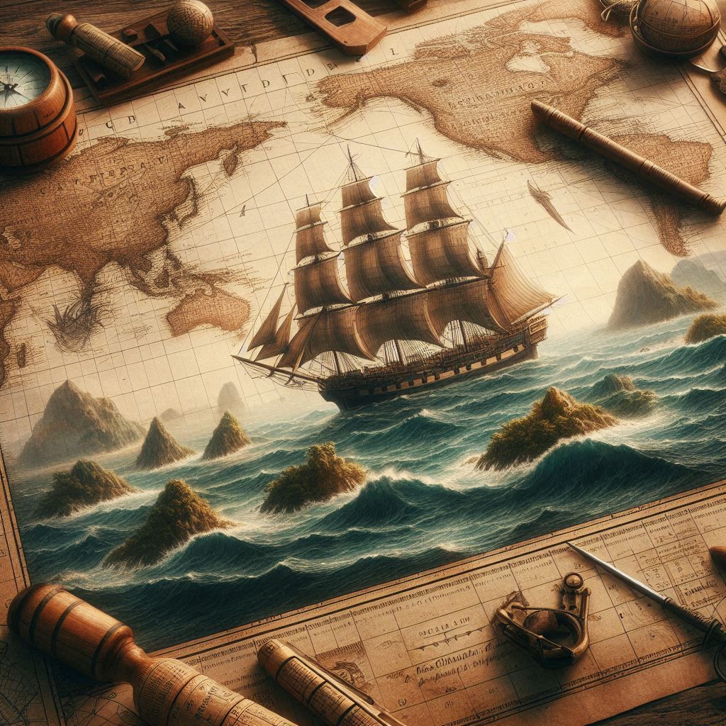
In the early 19TH century, a young and curious Charles Darwin set out on a journey that would transform the way humanity understood life on earth. His voyage aboard the HMS Beagle was not only an expedition across oceans but also an odyssey of discovery into intricate processes shaping the natural world.
His voyage aboard the HMS Beagle was not only an expedition across oceans but also an odyssey of discovery into intricate processes shaping the natural world.
Why did Darwin take the trip?
In 1831, Darwin, then a 22-year-old aspiring naturalist, received an unexpected invitation to join the crew of the HMS Beagle. The ship was tasked with a five-year mission to chart the coastlines of South America and update nautical maps. Darwin was recommended for the position of a naturalist, someone who could collect specimens and make observations about the natural world. Though his father initially opposed the idea, Darwin’s passion for nature and exploration won out. Little did anyone know; this trip would provide the foundation for a ground breaking scientific theory.
The journey was not just adventure- it was an opportunity for Darwin to explore the world’s diversity of life, something that had long intrigued him. The HMS Beagle set sail from England on December 27,1831, stopping at various locations across the southern hemisphere, including South America, Africa, and Australia. The true turning point of the voyage, however, came when the ship arrived at the Galapagos Islands.
Where are the Galapagos Islands?
The Galapagos Islands are a remote archipelago (a group of islands surrounded by sea) located in the Pacific Ocean, about 600 miles west of the coast of Ecuador. The islands are volcanic in origin and are home to a variety of unique plant and animal species. Isolated from the mainland, the Galapagos provided Darwin with a living laboratory for studying life in a way that had not been possible before.
When Darwin arrived at the Galapagos in September 1835, he was stuck by the peculiarities of the islands’ wildlife. Each island seemed to host its own distinct species, similar to but subtly different from those on other islands. These differences hinted at a deeper story of adaptation and evolution. Apparently, this isolation of species on different islands allowed them to evolve independently, resulting in striking variations from their mainland counterparts.
Darwin’s observation in the Galapagos
The finches of the Galápagos were among Darwin’s most famous discoveries. Though not appreciated initially, he soon realized that each island hosted finches with distinct beak shapes and sizes, perfectly adapted to the food available in their specific environments. For example, some finches had large, robust beaks ideal for cracking seeds, while others had slender, pointed beaks suited for eating insects. These variations led Darwin to wonder: Could these birds have originated from a common ancestor and diversified to survive in different conditions?
The islands are volcanic in origin and are home to a variety of unique plant and animal species. Isolated from the mainland, the Galapagos provided Darwin with a living laboratory for studying life in a way that had not been possible before.
The giant tortoises of the Galápagos also captivated Darwin. On one island, the tortoises had long necks and saddle-shaped shells, which allowed them to reach vegetation high off the ground. On another island, the tortoises had dome-shaped shells and shorter necks, suited for feeding on low-lying plants. These adaptations seemed to be perfectly tailored to the environment of each island, suggesting that the tortoises had evolved to fit their habitats.
As Darwin’s observations extended beyond the Galápagos, noticed striking similarities between species on different continents. For example, he observed the rhea, a large, flightless bird from South America, and compared it to the emu in Australia and the ostrich in Africa. Though these birds lived on different continents, they shared many similarities—large bodies, strong legs, and the inability to fly. This was a classic example of convergent evolution, a process in which unrelated species evolve similar traits because they occupy similar ecological niches. The rhea, emu, and ostrich had all evolved to thrive in wide-open spaces, but from different ancestral roots.
In South America, he encountered fossils of extinct animals, such as Megatherium (a giant ground sloth) and Glyptodon (a massive armadillo-like creature). These fossils bore striking resemblances to living species in the same region, hinting at a gradual change over time rather than the sudden, miraculous creation of life.
The Theory of Natural Selection
Darwin’s experiences and observations during the voyage led him to formulate his theory of evolution by natural selection. He theorized that in any population, there are natural variations between individuals. Some of these variations provide an advantage in survival and reproduction in given natural surroundings, allowing those traits to be passed down to future generations. Over time, these small changes could accumulate and lead to the emergence of a new species.
In the case of the Galápagos finches, for instance, birds with the different beak shapes survived in different islands because they were better suited to survive and reproduce in the environment provided by that particular island. This process of natural selection ensured that advantageous traits became more common over generations, eventually leading to the formation of distinct species.
Darwin, however, contradicted Lamarck’s ideas. He argued that various traits naturally occurred in the nature that were inherited through generations, (Darwin’s biggest lacuna is that he did not know how variation occurred. He was not familiar with genetics and genes) not through effort or use.
Contrasts with Lamarck and Other Theories
Darwin’s ideas were not entirely unprecedented. As earlier stated, before him, Jean-Baptiste Lamarck had proposed a theory of evolution based on the inheritance of acquired characteristics. Lamarck believed that organisms could change during their lifetimes and pass these changes on to their offspring to survive in the environment they lived in. For example, he argued that giraffes developed long necks because their ancestors stretched their necks to reach high leaves, and this trait was inherited by the next generation.
Darwin, however, contradicted Lamarck’s ideas. He argued that various traits naturally occurred in the nature that were inherited through generations, (Darwin’s biggest lacuna is that he did not know how variation occurred. He was not familiar with genetics and genes) not through effort or use. In his view, giraffes with naturally longer necks had a survival advantage over those who did not have them in the region which harboured long trees, enabling them to access food which those with shorter necks would not be able to access. Over generations, this advantage would lead to a population dominated by long-necked giraffes. It was the adaptive significance of the long neck variant that would the fittest for survival in that region.
Darwin also challenged other theories prevailing- at that time, such as special creation, which held that species were created in their present form by divine intervention. He disagreed with Georges Cuvier’s catastrophism, a theory championed by, which suggested that species were periodically wiped out by catastrophic events, and new ones were created afterward. Instead, Darwin argued that species evolved gradually over time in response to changes in their environments.
The Impact of Darwin’s Work
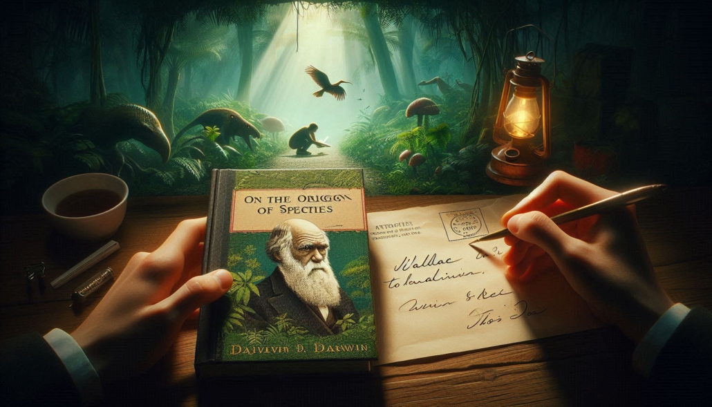
After years of refining his ideas and gathering more evidence during his voyage, he was initially hesitant to publish his ideas. The theory of natural selection was so radical that he feared the backlash it would provoke. However, situation took dramatic turn when in1858, another British naturalist, working in South America and Asia, Alfred Russel Wallace, sent Darwin a manuscript in which he propounded the concept of evolution quite similar to what Darwin had been gathering evidence to demonstrate. He had sent it to Darwin for his comments. Darwin showed this correspondence to his friends and conceded that he had been beaten by Wallace in this race. However, his friends, Hooker and Charles Lyell succeeded in persuading him to present a joint report with Wallace to the Linnean Society of London. Later, Darwin published his book-On the Origin of Specie in 1859, in which- he presented a compelling argument for the theory of evolution by natural selection, drawing on his extensive observations of animals like finches, tortoises, and rheas, as well as fossil evidence. The book forever changed the course of biology.
However, his friends, Hooker and Charles Lyell succeeded in persuading him to present a joint report with Wallace to the Linnean Society of London. Later, Darwin published his book-On the Origin of Specie in 1859
At first, the scientific community was divided, but over time, more and more evidence supported Darwin’s theory. Advances in genetics and palaeontology confirmed that species evolve through gradual changes, driven by natural selection. Today, Darwin’s theory is the foundation of modern biology, and his voyage aboard the HMS Beagle remains one of the most significant journeys of discovery in the history of science.
Darwin’s observations didn’t just map the world’s coastlines they mapped the history of life on Earth. Through his careful studies of the animals, plants, and fossils he encountered, Darwin uncovered the process that drives the evolution of species. Natural selection, he realized, was the key to the understanding of how life changes and adapts. There were no sudden, miraculous events that shaped life, but a slow, continuous process of change, where small differences in traits cumulatively determined survival, and where life evolved in response to the environment. The Galápagos Islands, a living laboratory for evolution, remain a symbol of Darwin’s legacy and the power of curiosity to unlock the mysteries of the natural world.
Darwin’s story reminds us that the great race of life has no finish line and the world is not static; it is ever-changing, shaped by the forces of adaptation and survival. His voyage was not just a journey of discovery but a revelation of the intricate processes that define life itself. The great race of life continues, a testament to the power of chance, variation, and the enduring adaptability of life.
Key Concepts to Remember:
Natural Selection: The process by which species adapt to their environment through survival and reproduction of the fittest.
Adaptation: The process by which an organism becomes better suited to its environment.
Evolution: The gradual change of species over time.
Common Ancestor: The concept that different species share a common evolutionary origin.
Lamarckism vs. Darwinism: Lamarck believed in inheritance of acquired characteristics (e.g., giraffes stretching their necks), while Darwin emphasized the role of natural selection and inherited traits that were advantageous from birth.
Catastrophism (Cuvier): The idea that species were wiped out by sudden, catastrophic events, as opposed to Darwin’s view of gradual change over time.
Convergent Evolution: The idea that similar ecological niches can lead to similar traits developing in different species (e.g., the similar traits of the rhea, emu, and ostrich).
https://curiosciencequest.com/wp-content/uploads/2025/04/ntural_selection_1-e1745933420407.png4101120info@curiosciencequest.comhttp://curiosciencequest.com/wp-content/uploads/2025/03/Untitled_design__1_-removebg-preview-1-300x300.pnginfo@curiosciencequest.com2025-04-29 13:43:442026-09-11 18:18:57Natural Selection Explained: Darwin, Wallace, Lamarck & How Evolution Works