 curio science quest
Part 2:The First Clues of Cell Division
A Journey from Cork to Chromosomes
The Secret of Mitosis: How Scientists Discovered Cell Division
By the mid-1800s, the microscope had become the scientist’s window into life. But looking inside a living cell was still very difficult. Cells were tiny, transparent, and constantly moving.
To solve this, scientists began experimenting with dyes and stains. Coloured chemicals could bind to certain parts of a cell, making invisible structures visible. This simple trick changed biology.
Carmine and the First Threads
One of the most useful dyes was carmine, a deep red pigment extracted from insects. When applied to cells, it stained the dense material inside the nucleus more strongly than the rest of the cell.
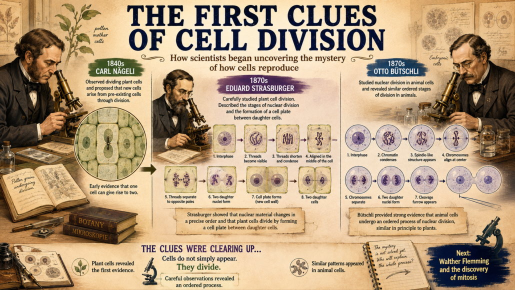
In the 1840s and 1850s, botanists such as Carl Nageli and Hugo von Mohl began to describe “thread-like” forms in plant cell nuclei. They didn’t fully understand what they were looking at, but their drawings showed thin, elongated fibers that seemed to appear during cell division.
These threads were, in fact, chromosomes though that word did not yet exist.

Figure 1: Early drawings by Nageli showing thread-like structures in dividing plant cells. (cartoon)
Robert Remak and Rudolf Virchow
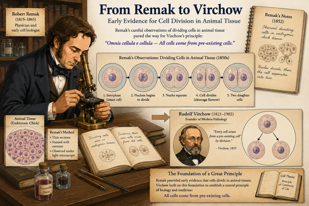
Meanwhile, in 1841, Robert Remak, a young German physician, was studying the development of chicken embryos. Using careful observation and early staining, he noticed that new cells did not appear spontaneously. Instead, they formed by binary division that is one cell splitting into two.
This was one of the earliest direct confirmations of Virchow’s later principle: cells arise from cells.
But Remak’s work was not widely accepted at first. It was Virchow, a more influential figure, who popularized the idea in 1855 with his famous phrase “Omnis cellula e cellula.”
Still, Remak deserves credit for being among the first to carefully document cell division in animal tissue.
Strasburger and the Plants
By the 1870s, techniques had advanced further. German botanist Eduard Strasburger used stronger microscopes and refined stains to study plant cells, especially in lilies and algae.
He provided detailed descriptions of the nucleus dividing into two during cell reproduction. He also noticed that the thread-like forms which we now recognize as chromosomes moved in a regular, organized way.
Strasburger’s observations hinted at a precise, step-by-step process. Division was not random. It followed an orderly sequence.

Figure 3 By the 1870s, Strasburger’s refined plant studies and Bütschli’s animal research revealed the universal ‘threads’ of life early glimpses of what we now call chromosomes.
Butschli and the “Nuclear Threads”
Another important contributor was Otto Butschli, who in the 1870s studied the eggs of worms and other simple animals. His work provided evidence that the “threads” were universal not just in plants, but in animals too.
This was a crucial step. The mysterious fibers inside cells were not accidents or artifacts of staining. They were real biological structures, present in all dividing cells.
By now, scientists were seeing glimpses of chromosomes, though they did not yet understand their true importance.
The Stage Is Set
By the late 19th century, one thing was clear: cells divided by a process involving the nucleus. Inside the nucleus, thread-like structures appeared, lined up, separated, and disappeared again.
But who would finally put the pieces together into a complete, step-by-step description?
That task would fall to a German anatomist named Walther Flemming.
Part 3: Walther Flemming and the Birth of “Mitosis”, we will explore how Flemming, using aniline dyes and patient observation, became the first person to clearly describe the entire process of mitosis. Continue the journey…
 curio science quest
 curio science quest
 curio science quest
  curio science quest
 Curio Science Quest
 curio science quest
  curio science quest
Leave a Reply
Want to join the discussion?Feel free to comment!