Tag Archive for: history of cell biology

Part 4: The story of chromosomes and inheritance

A Journey from Cork to Chromosomes

The Secret of Mitosis: How Scientists Discovered Cell Division


Chromosomes and the Secret of Inheritance

Walther Flemming had shown the precise and orderly sequence of cell division. But what were those mysterious threads? Why did they divide so carefully? And what did they carry?

The answers would come from a new generation of scientists at the turn of the 20th century.


Chromosomes Get Their Name

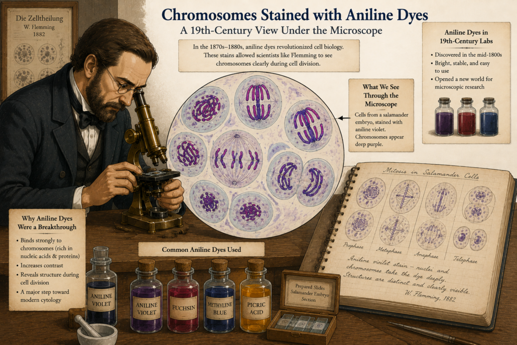
In 1888, a German anatomist named Heinrich Wilhelm Waldeyer gave the threads a new name: chromosomes.

  • “Chromo” means colour.
  • “Soma” means body.

Why? Because they picked up colour so vividly with the new dyes. The name stuck, and chromosomes became the stars of cell biology.

A Journey from Cork to Chromosomes
Figure 1: Chromosomes stained vividly with aniline dyes under a 19th-century microscope. (cartoon)

From Mitosis to Inheritance

Scientists now knew that chromosomes split evenly during mitosis. That meant daughter cells always received the same number. But another big mystery remained: How were traits passed from parents to offspring?

Gregor Mendel had already published his famous pea plant experiments back in 1866, showing that traits followed mathematical rules of inheritance.
Yet his work was forgotten for decades.

When it was rediscovered around 1900, scientists suddenly had two puzzles to connect:

  1. Mendel’s invisible units of heredity.
  2. The visible chromosomes that divided with such precision.

Could they be the same thing?


Theodor Boveri: A Careful Experimenter

The German biologist Theodor Boveri (1862–1915) was one of the first to see the connection.

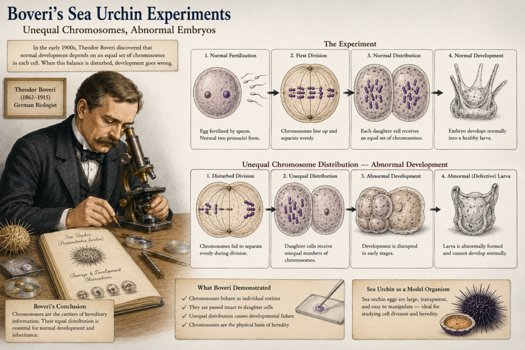
Working with sea urchin eggs, he performed delicate experiments. Sea urchins were perfect because their eggs were transparent, large, and divided rapidly. Boveri discovered something striking:

  • A cell could not develop properly unless it received a complete set of chromosomes.
  • If the distribution was abnormal if too many or too few chromosomes went into a cell, development failed.

This meant chromosomes were not just visible threads. They carried essential information needed for life.

A Journey from Cork to Chromosomes
Figure 2: Boveri’s sea urchin experiments — showing abnormal embryos when chromosomes were unevenly distributed. (cartoon)

Chromosomes as the Carriers of Heredity

Boveri concluded: chromosomes must be the physical carriers of heredity. Every single one mattered.
He was cautious but confident but the chromosomes were not identical; each had unique information. This was revolutionary. He had taken Flemming’s careful descriptions and added a powerful idea: chromosomes were not just for cell division, but also for inheritance of traits.


Walter Sutton and the Chromosome Theory

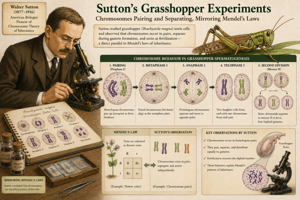
At the same time, across the Atlantic, a young American scientist named Walter Sutton was studying grasshopper cells. In 1902, Sutton noticed that chromosomes came in pairs. During cell division, each pair separated, with one chromosome going to each new cell. He realized this matched Mendel’s laws of inheritance:

  • Each parent contributes one factor (gene).
  • The factors separate and combine again in offspring.

Chromosomes behaved exactly like Mendel’s “hereditary units.”

A Journey from cork to chromosomes
Figure 3: Sutton’s grasshopper chromosomes pairing and separating, mirroring Mendel’s laws. (cartoon)

The Chromosome Theory of Inheritance

Together, Boveri and Sutton laid the foundation for the chromosome theory of inheritance.

It said:

  • Genes are located on chromosomes.
  • Chromosomes are passed from cell to cell, and from parent to offspring, through mitosis and meiosis.

This was the missing link between cell biology and genetics.


Why This Was a Turning Point

Up until Flemming, mitosis was just a strange cellular dance. With Boveri and Sutton, the meaning became clear:

  • Chromosomes were the carriers of heredity.
  • Mitosis ensured that every new cell received the full library of instructions.
  • Life’s continuity, from one cell to another, depended on this precise mechanism.

The mystery of cell division had transformed into the foundation of modern genetics.


A Legacy That Still Shapes Science

Today, we know chromosomes are made of DNA tightly wrapped around proteins.
We know they carry thousands of genes that code for every trait in living organisms.
But that knowledge rests on the careful experiments of Flemming, Boveri, Sutton, and their colleagues.

They took what seemed like meaningless threads and revealed them as the very carriers of life’s code.

A Journey from Cork to Chromosomes
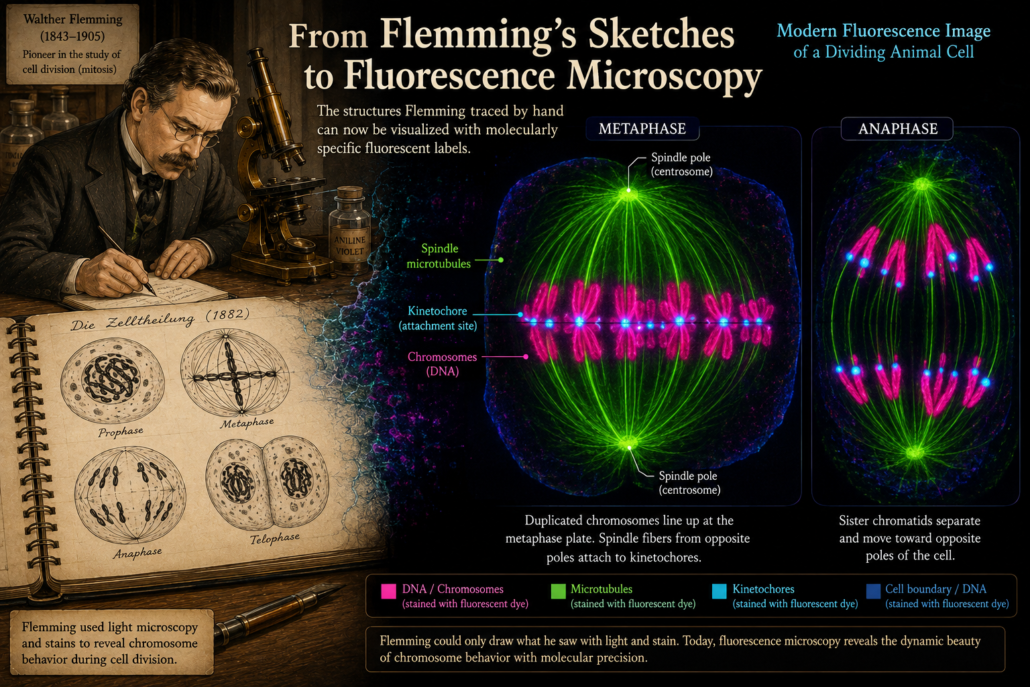
Figure 4: Modern fluorescence image of dividing chromosomes echoing Flemming’s hand-drawn sketches. (cartoon)

Part 5: Why Mitosis Matters Today, we will explore how this history connects to modern science: from cancer research to cloning, stem cells, and genetic medicine. Continue the journey…

Part 5: Why Mitosis Matters.
Let, connect the history to modern discoveries and why cell division still holds so many secrets.

Part 2:The First Clues of Cell Division

A Journey from Cork to Chromosomes


The Secret of Mitosis: How Scientists Discovered Cell Division

By the mid-1800s, the microscope had become the scientist’s window into life. But looking inside a living cell was still very difficult. Cells were tiny, transparent, and constantly moving.

To solve this, scientists began experimenting with dyes and stains. Coloured chemicals could bind to certain parts of a cell, making invisible structures visible. This simple trick changed biology.


Carmine and the First Threads

One of the most useful dyes was carmine, a deep red pigment extracted from insects. When applied to cells, it stained the dense material inside the nucleus more strongly than the rest of the cell.

In the 1840s and 1850s, botanists such as Carl Nageli and Hugo von Mohl began to describe “thread-like” forms in plant cell nuclei. They didn’t fully understand what they were looking at, but their drawings showed thin, elongated fibers that seemed to appear during cell division.

These threads were, in fact, chromosomes though that word did not yet exist.


 The Secret of Mitosis: How Scientists Discovered Cell Division

Figure 1: Early drawings by Nageli showing thread-like structures in dividing plant cells. (cartoon)


Robert Remak and Rudolf Virchow

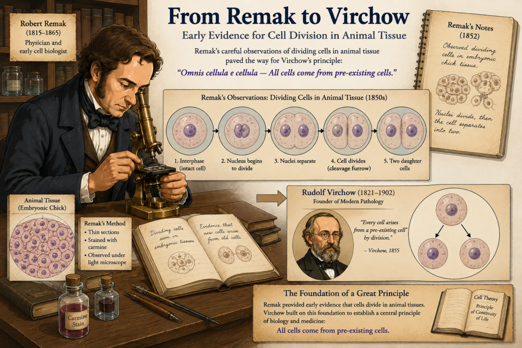
Meanwhile, in 1841, Robert Remak, a young German physician, was studying the development of chicken embryos. Using careful observation and early staining, he noticed that new cells did not appear spontaneously. Instead, they formed by binary division that is one cell splitting into two.

This was one of the earliest direct confirmations of Virchow’s later principle: cells arise from cells.

But Remak’s work was not widely accepted at first. It was Virchow, a more influential figure, who popularized the idea in 1855 with his famous phrase “Omnis cellula e cellula.”

Still, Remak deserves credit for being among the first to carefully document cell division in animal tissue.


A Journey from Cork to Chromosomes
Figure 2: Early evidence for cell division in animal tissue Remak’s careful work paved the way for Virchow’s principle that all cells come from pre-existing cells.

Strasburger and the Plants

By the 1870s, techniques had advanced further. German botanist Eduard Strasburger used stronger microscopes and refined stains to study plant cells, especially in lilies and algae.

He provided detailed descriptions of the nucleus dividing into two during cell reproduction. He also noticed that the thread-like forms which we now recognize as chromosomes moved in a regular, organized way.

Strasburger’s observations hinted at a precise, step-by-step process. Division was not random. It followed an orderly sequence.


The Secret of Mitosis: How Scientists Discovered Cell Division

Figure 3 By the 1870s, Strasburger’s refined plant studies and Bütschli’s animal research revealed the universal ‘threads’ of life early glimpses of what we now call chromosomes.


Butschli and the “Nuclear Threads”

Another important contributor was Otto Butschli, who in the 1870s studied the eggs of worms and other simple animals. His work provided evidence that the “threads” were universal not just in plants, but in animals too.

This was a crucial step. The mysterious fibers inside cells were not accidents or artifacts of staining. They were real biological structures, present in all dividing cells.

By now, scientists were seeing glimpses of chromosomes, though they did not yet understand their true importance.


The Stage Is Set

By the late 19th century, one thing was clear: cells divided by a process involving the nucleus. Inside the nucleus, thread-like structures appeared, lined up, separated, and disappeared again.

But who would finally put the pieces together into a complete, step-by-step description?

That task would fall to a German anatomist named Walther Flemming.


 Part 3: Walther Flemming and the Birth of “Mitosis”, we will explore how Flemming, using aniline dyes and patient observation, became the first person to clearly describe the entire process of mitosis. Continue the journey…


Part 1: The Birth of Cell Theory

A Journey from Cork to Chromosomes

History of Cell Biology: From Hooke’s Cork Cells to Chromosomes


From Cork to Cells: Robert Hooke and the Birth of Cell Biology

What is the history of cell biology?

The history of cell biology began with Robert Hooke’s observation of cork cells in 1665 and progressed through the discoveries of Leeuwenhoek, Brown, Schleiden, schwann, Virchow and Flemming.


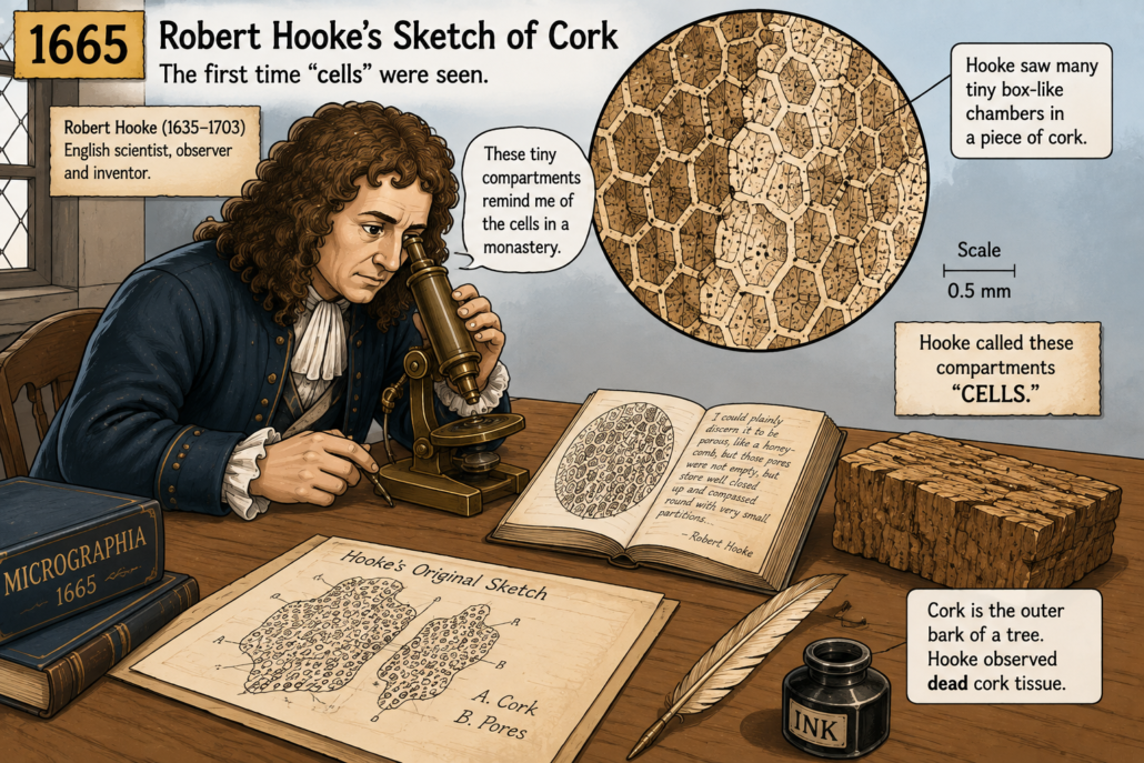
Imagine peeling the bark from a tree and placing a thin slice under a primitive microscope. That is exactly what happened in 1665, when the English scientist Robert Hooke looked at cork. Through his hand-built compound microscope, he saw something no one had described before: tiny, box-like compartments.

Hooke thought they looked like the small rooms where monks lived. He called them “cells.”

This was the first recorded use of the word “cell” in biology. Of course, Hooke was not actually seeing living cells. He was looking at the empty walls of dead plant tissue. The living contents had long dried away, leaving behind only the rigid outlines.

But his observation was revolutionary. Hooke’s book Micrographia (1665) inspired curiosity across Europe. The idea that nature could be studied with magnifying lenses became a serious pursuit.


A Journey from Cork to Chromosomes
Figure 1: Robert Hooke’s 1665 sketch of cork, showing tiny “cells.”showing tiny “cells.”

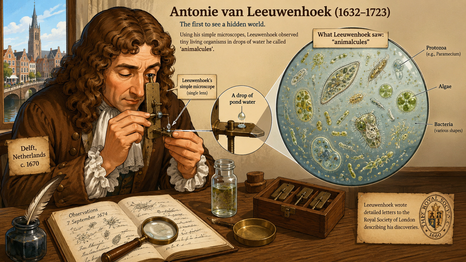
At the same time, another pioneer was grinding glass into lenses that were far better than Hooke’s. This man was Antonie van Leeuwenhoek, a Dutch tradesman with a passion for magnification. His microscopes were small, single-lens devices, but they were powerful

Leeuwenhoek used his lenses to look at everything he could imagine from pond water, blood, semen, even scrapings from his own teeth. What he discovered amazed the world. He described tiny moving creatures that no one had seen before and named them ‘animalcules.

These were the first recorded observations of living microorganisms, bacteria, protozoa, and even sperm cells.


History of Cell Biology: From Hooke’s Cork Cells to Chromosomes
Figure 2: Antonie van Leeuwenhoek’s simple microscope and his “animalcules” in water droplets.

For the first time, people began to understand that life was built from tiny, hidden structures. But there was still no single theory to explain it all. Hooke had described cells in plants, and Leeuwenhoek had observed animal and bacterial forms. The connection between them remained unclear.

More than 150 years would pass before scientists connected these dots.


The Nucleus Appears

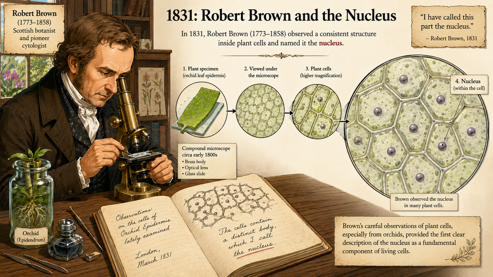
In 1831, Scottish botanist Robert Brown was studying orchids. While observing the cells of these plants, he noticed a consistent, dark, rounded body inside each one. He named it the nucleus.

This was a turning point. Cells were not just empty spaces surrounded by walls. They had internal parts. They had organization. The nucleus seemed to be a central structure, and later it would prove essential for heredity.


A Journey from Cork to Chromosomes
Figure 3: Robert Brown’s 1831 drawing of plant cells, highlighting the nucleus.

The Cell Theory

By the 1830s and 1840s, microscopes were improving. Glassmaking had advanced, and staining methods with dyes like carmine and iodine made cellular details easier to see.

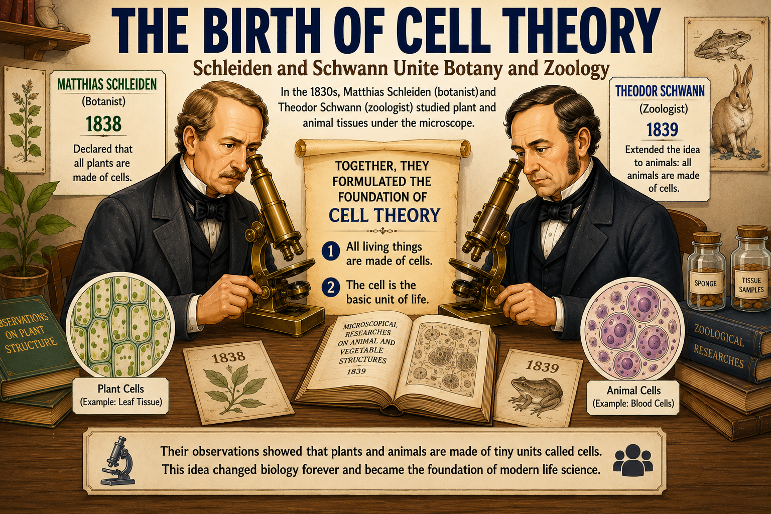
In Europe, two scientists named Matthias Schleiden (a botanist) and Theodor Schwann (a zoologist) began studying plant and animal tissues under the microscope.

In 1838, Schleiden declared that all plants are made of cells. In 1839, Schwann extended the idea to animals. Together, they laid down the foundation of what became known as the Cell Theory:

  1. All living things are made of cells.
  2. The cell is the basic unit of life

History of Cell Biology: From Hooke’s Cork Cells to Chromosomes
Figure 4: Schielden and Schwann unite Botany and Zoology

This was revolutionary. Suddenly, plants, animals, and even humans were united by a common building block.

Yet one mystery remained: Where do new cells come from?

At the time, many scientists still believed in “spontaneous generation” the idea that life could appear out of non-living matter. Some thought cells could simply crystallize or form a new inside organisms.

It was not until 1855 that German physician Rudolf Virchow delivered the famous phrase: “Omnis cellula e cellula.” Every cell comes from another cell.

This simple idea challenged the long-held belief that cells could arise by spontaneous generation. It showed cells, could not appear from nothing but they had to come from the division of pre-existing cells.

Virchow’s principle would become the third pillar of the Cell Theory.


A Hidden Process

But here lay the puzzle: How exactly does one cell become two?

Scientists had no clear picture of the process. They could sometimes glimpse strange, thread-like structures inside dividing cells, especially when they used new stains. But these observations were brief and confusing.

The stage was set for a discovery that would change biology forever the discovery of mitosis, the process of cell division.


Part 2: The First Clues of Cell Division, we will explore the 1800s when new dyes and microscopes revealed mysterious threads inside cells, and Virchow’s principle met its first experimental tests. Continue the journey…..

 The Secret of Mitosis: How Scientists Discovered Cell Division